Tadalafil appartiene alla classe degli inibitori selettivi della fosfodiesterasi di tipo 5, con un profilo farmacocinetico caratterizzato da un’emivita terminale di circa diciotto ore. Dopo somministrazione orale viene assorbito rapidamente e raggiunge concentrazioni plasmatiche massime in due ore. La biotrasformazione avviene principalmente tramite CYP3A4 con formazione di metaboliti inattivi, escreti in prevalenza con le feci. L’elevato legame alle proteine plasmatiche (>90%) assicura una distribuzione stabile. Nei confronti delle altre molecole della stessa classe, cialis compresse italia è noto per la durata prolungata dell’attività farmacologica.
Doi:10.1016/s0889-1591(03)00054-0
Brain,Behavior,and Immunity 17 (2003) 251–259
Acute stress evokes selective mobilization of T cells that
differ in chemokine receptor expression: a potential
pathway linking immunologic reactivity to
Jos A. Bosch,a Gary G. Berntson,b John T. Cacioppo,c Firdaus S. Dhabhar,d,e
a Periodontology Section, The Ohio State University, College of Dentistry, 305 West 12th Avenue, P.O. Box 182357,
b Department of Psychology, The Ohio State University, Columbus, OH, USA
c Department of Psychology, University of Chicago, Chicago, IL, USA
d Institute for Behavioral Medicine Research, The Ohio State University, Columbus, OH, USA
e Department of Oral Biology, The Ohio State University, College of Dentistry, Columbus, OH, USA
Received 31 October 2002; received in revised form 7 February 2003; accepted 19 February 2003
T lymphocytes and monocytes/macrophages are the most abundant cells found in the atherosclerotic plaque. These
cells can migrate towards the activated endothelium through the local release of chemotactic cytokines,or chemokines. Given the important role of leukocyte migration in atherosclerosis and the role of stress in mediating leukocyte traf-ficking,the present study examined the effects of an acute stressor on the redistribution of T cells (CD3+) andmonocytes that express the chemokine receptors CCR5,CCR6,CXCR1,CXCR2,CXCR3,and CXCR4. Forty-fourundergraduate students underwent a public speaking task. The acute stressor induced sympathetic cardiac activation,parasympathetic cardiac withdrawal,lymphocytosis,and monocytosis (all p < :001). Although the total number of Tlymphocytes did not change,there was a selective increase in the number of circulating T cells expressing CXCR2,CXCR3,and CCR5. The ligands of these receptors are chemokines known to be secreted by activated endothelial cells. Analyses of individual differences in stress-induced responses demonstrated a positive relationship between sympatheticcardiac reactivity and mobilization of the various T cell subsets (:35 < r < :56; p < :05). For the monocytes,all sub-populations increased in parallel with total monocyte numbers,with no relation to changes in sympathetic cardiacdrive. These results indicate that acute stress induces a mobilization of T cells that are primed to respond to inflamedendothelium. Acute stressors may thus promote the recruitment of circulating immune cells into the sub-endothelia,andtherefore accelerate atherosclerotic plaque formation and potentially contribute to the complications that follow acutestressful events. This mechanism may help explain the link between stress,reactivity,and cardiovascular disease. Ó 2003 Elsevier Science (USA). All rights reserved.
Keywords: Cardiovascular disease; Acute psychological stressor; Psychoneuroimmunology; Cardiovascular reactivity; Autonomicbalance; Lymphocyte redistribution; Chemokines
* Corresponding author. Fax: 1-614-292-4612.
0889-1591/03/$ - see front matter Ó 2003 Elsevier Science (USA). All rights reserved. doi:10.1016/S0889-1591(03)00054-0
J.A. Bosch et al. / Brain, Behavior, and Immunity 17 (2003) 251–259
Acute psychological stressors are known to modulate
this process of leukocyte trafficking and to enhance
Many lines of evidence,ranging from pathologic
subsequent cellular immune responses in the local tissues
analyses to epidemiological studies,show that athero-
(Dhabhar & McEwen,1997,1999; Dhabhar,Miller,
sclerosis is intrinsically an inflammatory disease (Libby,
Stein,McEwen,& Spencer,1994; Sanders & Straub,
Ridker,& Maseri,2002; Ross,1999). The initiation of
2002). If,as the exisiting evidence suggests,migratory
inflammatory reactions is a complex process involving
responses of leukocytes are crucial in the development of
the coordinated expression of cellular adhesion mole-
atherosclerotic lesions,then acute stress may influence
cules and chemotactic cytokines (chemokines),which
atherosclerotic plaque formation in part through its ef-
recruit blood-derived leukocytes to the site of inflam-
fects on leukocyte migration and recruitment.
mation. The recruitment of leukocytes by chemokines
The magnitude of cardiovascular system responses to
into the sub-endothelium of the vascular wall is a major
acute stressors (‘‘cardiovascular reactivity’’) is consid-
aspect of atherogenesis. T lymphocytes are among the
ered a potential risk factor for cardiovascular disease
first cells to infiltrate the sub-endothelium (Libby et al.,
progression and its acute clinical manifestations (Kop,
2002; Ross,1999; Song,Leung,& Schindler,2001),and
1999; Krantz,Kop,Santiago,& Gottdiener,1996; Ro-
remain a major local cell population throughout the
zanski,Blumenthal,& Kaplan,1999; Sheps et al.,2002).
atherosclerotic process. These T cells subsequently se-
Likewise,immune reactivity (the response of immune
crete cytokines (e.g.,interferon-c,TNF-a,and interleu-
parameters during acute stress) has been proposed as a
potential predictor for vulnerability to immune medi-
atherosclerotic response. Monocytes are another critical
ated disease (Cacioppo et al.,1998; Cohen et al.,2002;
constituent of the atherosclerotic response. Once resi-
Sanders & Straub,2002). Cardiovascular and immune
dent in the vessel wall,monocytes develop into macro-
reactivity are correlated phenomena that are both de-
phages as they take up oxidized low-density lipoprotein
termined by sympathetic nervous system activation
and differentiate into so-called foam cells. Macrophages
(Cacioppo et al.,1995; Sgoutas-Emch et al.,1994;
and lipid-laden foam cells are implicated as prime cul-
Uchino,Cacioppo,Malarkey,& Glaser,1995). Thus,it
prits in the events that ultimately complicate athero-
is possible that the observed link between sympathetic
sclerosis (Libby et al.,2002; Ross,1999).
cardiac reactivity and cardiovascular disease manifesta-
It is likely that the endothelium itself initiates this
tions is mediated in part through immunological path-
process of leukocyte recruitment (Libby et al.,2002;
ways. The present study examined the effects of an acute
Reape & Groot,1999; Shin,Szuba,& Rockson,2002).
stressor on the redistribution of T cells (CD3+) and
Endothelial cells can secrete numerous chemokines
monocytes that express the chemokine receptors CCR5,
upon activation by molecules derived from the circula-
tion and adjacent cells (e.g.,Burke-Gaffney,Brooks,&
findings suggest that immune reactivity,or at least some
Bogle,2002; Kotani,Hori,Matsumura,& Uchiyama,
aspects of this phenomenon,may also be relevant to the
2002; Mach et al.,1999; Qi & Kreutzer,1995; Seeger
development of cardiovascular disease.
et al.,2002). In fact,virtually all cardiovascular riskfactors (e.g.,increased LDL levels,hypertension,dia-betes,obesity,and infection) are capable of promoting
an inflammatory response in endothelial cells with theconcomitant secretion of inflammatory mediators (Lib-
by et al.,2002). Chemokines secreted by endothelialcells include Growth Regulated Oncogene (GRO,which
Forty-four university undergraduates (mean age 20,
has an a; b,and c sub-type),Epithelial Neutrophil Ac-
range 18–27 years,22 male) volunteered to participate in
tivating peptide-78 (ENA-78),Neutrophil Activating
this study as part of a longitudinal study on psychoso-
Protein-2 (NAP-2),and Interleukin 8 (IL-8). These
cial factors and wound healing. Participants gave writ-
chemokines are all ligands for the pleiotrophic chemo-
kine receptor CXCR2,whereas IL-8 can also stimulate
compensation for their participation. Participants were
the chemokine receptor CXCR1. Other examples of
ineligible if they were using medication,or reported
chemokines secreted by endothelial cells are Interferon-
health problems indicative of cardiovascular,inflam-
c Inducible Protein-10 (IP-10),which binds to the
chemokine receptor CXCR3,and Regulated on Acti-vation Normal T cell Expressed and Secreted (RAN-
TES),which is a ligand for several chemokine receptorsincluding CCR5 (Burke-Gaffney et al.,2002; Oppen-
In preparation for the study,participants were in-
heim,Zachariae,& Goetzl,2000; Wang,Su,Gong,&
structed not to engage in strenuous physical exercise,
and to refrain from using alcohol or non-prescription
J.A. Bosch et al. / Brain, Behavior, and Immunity 17 (2003) 251–259
drugs 24 h before the experimental sessions. In addition,
ECG R-wave across 1-min periods. From these 1-min
participants were instructed to abstain from smoking
ensembles,average levels were computed for heart rate
and caffeine the day of the experiment,to eat breakfast
(HR) and preejection period (PEP). These minute-by-
before 10 a.m. and stay null per os (except for water)
minute means were averaged over a 6-min pretask
from 10 a.m. onwards. Women were scheduled in the
baseline and over each 6-min stressor.
first week after their menses. Upon arriving at the
Interbeat intervals (IBI) were checked and edited for
General Clinical Research Center at 12:00 p.m.; (a) in-
artifacts by a detection algorithm developed by Berntson,
formed consent was obtained; (b) a 19-G indwelling
Quigley,Jang,and Boysen,1990. Respiratory sinus ar-
catheter was placed into the antecubital vein of the non-
rhythmia (RSA) was derived by the method of Porges
dominant arm; (c) participants were served a standard-
(Porges & Bohrer,1990),using the MXedit program
ized lunch (300 kcal,containing 10 g of protein,12 g of
(Delta Biometrics,Bethesda MD). This program converts
carbohydrate,and 16 g of fat) and water ad libitum; and,
the heart period series into a time series,applies a moving
(d) electrodes for electrocardiography (ECG) and im-
polynomial filter (polynomial ¼ 3,coefficients ¼ 21) to
pedance cardiography (ICG) were attached. Subse-
remove slow non-stationarities in the data,applies a
quently,while seated in supine position,participants
band-pass filter (.12–.40 Hz) to the residual series,and
filled out questionnaires and engaged in leisure reading.
then derives the natural log of the band variance. This
At 1:30 p.m.,a baseline blood sample was obtained and
corresponds to the statistical variance of the time sampled
the procedure for the laboratory stressor was initiated.
heart period data within the respiratory frequency band.
Changes in PEP were used to index changes in car-
diac sympathetic drive,whereas RSA was used to indexchanges in cardiac vagal tone.
The stress task consisted of two back-to-back spee-
ches,each with 2 min of preparation and 4 min of speech
delivery. To enhance social stress,the speeches werevideotaped and attended by a small audience consisting
Whole blood was collected into sodium heparin
of a female nurse,a female research assistant,and a male
tubes,and maintained at room temperature. Samples
psychologist. For the first speech,the participant had to
were prepared within 2 h after collection. White blood
defend him/herself after being falsely accused of shop-
cell counts were obtained on a hematology analyzer
lifting (Saab,Matthews,Stoney,& McDonald,1989),
(F800,Sysmex,McGraw Park,IL). Specific leukocyte
and for the second speech the participant gave a pre-
sub-types were identified by immunofluorescent anti-
sentation about his or her best and worst personal
body staining using flow cytometry (FACSCalibur,
characteristics (van Eck,Nicolson,Berkhof,& Sulon,
Becton–Dickinson,San Jose,CA). Whole blood was
1996). In order to standardize timing and instructions
stained using phycoerythrin (PE) and FITC conjugated
for the task,the instructions were presented on a video
monoclonal antibodies for chemokine receptors,and
screen. Including instructions,the task took 15 min. A
cychrome (CyChr) for CD3 (Pharmingen,San Diego,
second blood sample was obtained during second min-
CA). Briefly,cell suspensions were incubated with anti-
ute of the second presentation (13 min after initiation of
body for 20 min at room temperature,lysed with FACS
the task). Cardiac activity was recorded continuously.
Brand Lysing Solution (Becton–Dickinson,San Jose,CA),which results in a simultaneous lysis of red blood
cells and partial fixation of leukocytes,washed withPBS,and read on the FACSCalibur with 3000–5000
Assessment of cardiovascular responses focused on
events being acquired from each preparation. Matched
cardiac sympathetic and vagal control (Berntson,Caci-
antibody isotype controls were used to set negative
oppo,& Quigley,1993). Indices of sympathetic and
staining criteria. Data were analyzed using Cell Quest-
parasympathetic drive were obtained by analysis of
pro software (Becton–Dickinson,San Jose,CA).
ECG and thoracic impedance (ICG) signals (Berntson etal.,1997; Sherwood et al.,1990). The thoracic imped-
ance and ECG signals were recorded from six Ag/AgClspot-electrodes (AMI type 1650-005,Medtronic) using
the Vrije Universiteit Ambulatory Monitoring System
ducted to examine the effects of the laboratory stressor
(VU-AMS) device. Reliability and validity of the VU-
on mood,cardiovascular parameters,and cellular dis-
AMS device have been reported elsewhere (de Geus,
tribution. The ECG recording of one subject could not
Willemsen,Klaver,& van Doornen,1995; de Geus &
be completed due to a technical problem. For two sub-
van Doornen,1996; Willemsen,De Geus,Klaver,Van
jects,flow cytometry data was incomplete due to in-
Doornen,& Carroll,1996). The ECG and ICG com-
complete lysis of a blood sample. Degrees of freedom
plexes were ensemble averaged with reference to the
J.A. Bosch et al. / Brain, Behavior, and Immunity 17 (2003) 251–259
numbers of CD3+CXCR2+,CD3+CXCR3+,andCD3+CCR5+ lymphocytes (see Table 2). Analyses of
3.1. Psychological and cardiovascular reactions
these sub-populations expressed as% of total T cellnumbers yielded a similar pattern of results,showing a
Analysis of POMS subscales indicated that the
selective increase of CXCR2+,CXCR3+,and CCR5+
speech tasks were perceived as stressful,as evidenced by
T-cells within the total circulating T-cell pool (all
increases in tension-anxiety (Mbaseline 3.1 (SEM 0.5), Mtask
Fð43Þ > 6:5; p < :01). This analysis further revealed a
9.3 (SEM 0.8), Fð43Þ ¼ 21:34; p < :001) and anger-hos-
small decrease in CCR6+ T-cells relative to the total
tility (Mbaseline 0.18 (SEM 0.3), Mtask 2.3 (SEM 0.6),
number of T cells (Fð43Þ ¼ 6:18; p < :05).
Fð43Þ ¼ 10:73; p < :01). Replicating prior research,anal-
Data and statistical analyses for monocytes are pre-
yses confirmed that the acute psychosocial stressor ele-
sented in Table 3. The speaking stressor increased total
vated HR,increased cardiac sympathetic activation,and
monocyte numbers. A general increase was also seen for
produced vagal withdrawal (see Table 1). Because gen-
the various monocyte sub-populations,which paralleled
der and body composition (i.e.,BMI) have been re-
the increase in total monocyte numbers (see Table 3).
ported to moderate the autonomic responses to acute
That all monocyte subsets (as defined by chemokine
stressors (as well as being independent predictors of
receptor expression) changed to the same degree was
cardiovascular disease occurrence),we included these
further corroborated by non-significant changes in
potential modifiers in our statistical model. As shown in
monocyte subsets expressed as percentage of total
Table 1,controlling for these variables yielded compa-
rable results. Further analyses did neither reveal modi-
We again performed analyses controlling for possible
fying effects of age or smoking on cardiac responses.
moderating effects of gender and BMI. As shown inTables 2 and 3,these analyses yielded a similar pattern
of results. Subsequent exploratory analyses neither in-dicated moderating effects of smoking status or age.
Data and statistical analyses for the lymphocytes are
presented in Table 2. The acute stressor induced signif-
3.3. Associations between cellular and cardiac autonomic
icant lymphocytosis,whereas the total number of cir-
culating T lymphocytes did not increase. Furtheranalysis of T cell sub-population on basis of chemokine
To confirm the assumption that cardiac sympathetic
receptor expression,revealed significant increases in the
reactivity and immune reactivity are related phenomena,
Table 1Mean values (with SEM in parentheses) and results of statistical analyses (repeated measures ANOVA/ANCOVA) for cardiacautonomic measures during baseline and speech tasks
Table 2Mean values (with SEM in parentheses) and results of statistical analyses (repeated measures ANOVA/ANCOVA) for lymphocyte cellnumbers during baseline and speech tasks
J.A. Bosch et al. / Brain, Behavior, and Immunity 17 (2003) 251–259
Table 3Mean values (with SEM in parentheses) and results of statistical analyses (repeated measures ANOVA/ANCOVA) for monocyte cellnumbers during baseline and speech tasks
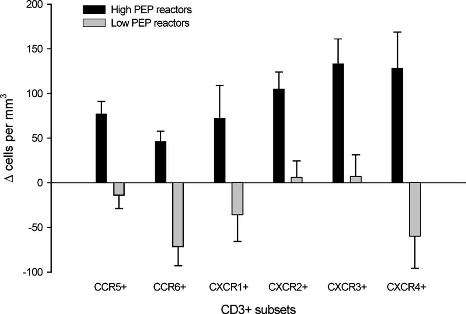
we compared cellular mobilization responses in subjects
low reactors exhibited no change or a decrease (see Fig.
that exhibited a high versus a low cardiac sympathetic
1). Independent t tests yielded significant differences for
reactivity. The cardiac PEP was utilized as a measure of
cardiac sympathetic drive. Reactivity was calculated as
(tð40Þ ¼ 3:62; p < :005),CD3+CXCR1+
ðPEPtask 1 þ PEPtask 2Þ=2 À PEPbaseline,and grouping into
p < :05),CD3+CXCR2+ (tð40Þ ¼ 3:72; p < :005),CD3+
high and low PEP reactors was based on a median split
CXCR3+ (tð40Þ ¼ 3:44; p < :005),and CD3+CXCR4+
of these reactivity scores. The average Dms for the high
(tð39Þ ¼ 3:47; p < :005) (see Fig. 1). Inspection of the
PEP reactors was )17.2 (SEM 1.8),and )1.5 (SEM 0.6)
scatter plots and computation of bivariate correlations
for the low PEP reactors (tð41Þ8:29; p < :001). High and
indicated that the associations between sympathetic
low PEP reactors also differed in HR reactivity
cardiac drive and cellular mobilization showed a near
(tð41Þ2:47; p < :05; Dbpm 21.9 (SEM 2.9) vs. 13.3 (SEM
linear relationship (see Table 4 and Fig. 2).
1.8)),whereas the two groups did not differ in vagal
Previous studies have reported that individual dif-
ferences in HR reactivity,instead of PEP,may predict
Ideographic analyses showed a general increase in
immune reactions e.g. (Benschop et al.,1998; Larson,
circulating T cell subsets for high PEP reactors,whereas
Ader,& Moynihan,2001; Sgoutas-Emch et al.,1994),
Fig. 1. Change in T cell sub-set numbers for subjects that exhibited a high versus a low cardiac sympathetic response (indexed aschanges in PEP) during the speech stressors. Bars indicate mean,lines indicate standard error of mean.
J.A. Bosch et al. / Brain, Behavior, and Immunity 17 (2003) 251–259
Table 4Pearson correlation coefficients and SpearmanÕs rank-order correlation coefficients (between brackets) of the association between PEPreactivity and changes in T cell numbers
Fig. 2. Scatter plots showing the relation between PEP reactivity and mobilization of CCR5+ and CXCR2+ T cells.
although the sympathetic component of cardiac reac-
towards the activated endothelium by chemokines that
tivity appears to be superior to overall reactivity (Caci-
are initially secreted by the local endothelial cells. The
oppo,1994; Cacioppo et al.,1995). This is consistent
present study investigated the effects of an acute stressor
with the present analyses. HR reactivity yielded a com-
(public speaking) on the mobilization of T cells and
parable pattern of results as presented in Fig. 1,albeit
monocytes that express receptors for these chemotactic
showing less pronounced differences. Indeed,comparing
factors. Whereas the total number of circulating CD3+
the high and low HR reactivity groups yielded signifi-
lymphocytes did not change,the speaking stressor in-
cant group differences only for CD3+CCR5+ (tð40Þ ¼
duced an increase in CD3+ cells expressing receptors for
chemokines that are known to be secreted by activated
endothelium (GRO,NAP-2,ENA-78,IL-8,IP-10 for
comparisons of high and low vagal/parasympathetic
cardiac reactors (indexed by changes in RSA) yielded no
Gaffney et al.,2002; Kotani et al.,2002; Qi & Kreutzer,
statistical differences in regard to T cell responses (for all
1995; Seeger et al.,2002; Wang et al.,1998). No change
was seen in the number of T cells carrying receptors for
Similar analyses as described above (i.e.,dividing our
chemokines that are not secreted by inflamed endothe-
sample in high vs low reactors based on median differ-
lial cells (SDF-1 for CXCR4,LARC/MIP-3a for
ence scores of PEP,HR,or RSA) were performed to
CCR6).1 The various monocyte subsets (as defined by
explore monocyte responses in relation to cardiac and
chemokine receptor expression),on the other hand,did
autonomic reactions. In contrast to the T lymphocytes,
not show such specificity: all subsets increased in
monocyte responses showed no significant associations
parallel with total monocyte numbers. Thus,the re-
with PEP reactivity,heart rate reactivity or vagal reac-
sults indicate a selective mobilization of T cells that
tivity: for all t tests, t < 1:5 and p > :2.
are sensitive to the chemotactic signals of an inflamedendothelium.
1 Chemokine receptors are know to exhibit a remarkable
T lymphocytes and monocytes/macrophages are the
pleiotrophism,and therefore it cannot be excluded that CXCR4
most abundant cells found in the atherosclerotic plaque
and CCR6 may show some responsivity to chemokines that are
(Libby et al.,2002; Ross,1999). These cells are attracted
secreted by inflamed endothelial cells.
J.A. Bosch et al. / Brain, Behavior, and Immunity 17 (2003) 251–259
Ideographic analysis,comparing mobilization re-
vided by research on the role of stress-induced migratory
sponses in subjects exhibiting high versus low sympa-
responses in acute cardiovascular complications such as
thetic cardiac reactivity,confirmed and extended the
results of nomothetic analyses. In line with previous
T cells constitute a heterogeneous population,and it
research (Cacioppo et al.,1995; Uchino et al.,1995),it
is well established that the CD8+ T cells in particular are
confirmed our assumption that sympathetic cardiac re-
mobilized during acute stress in humans (Benschop et
activity and immune reactivity are closely related phe-
al.,1996; Sanders & Straub,2002). Hence,the selective
nomena. In particular,increases in T cell subsets were
increases observed in our study might reflect changes in
prominent in the high PEP reactors but were absent or
this CD8+ sub-population. Examination of the litera-
even reversed for the low PEP reactors. For example,
ture suggests that this may only provide a partial ex-
whereas the CD3+CCR6+ subset showed a trend to-
planation. For example,we found an increase in T cells
wards increased cell numbers in high PEP reactors
that are positive for the receptors CXCR3 and CCR5,
(p ¼ :08),a significant decrease (p < :01) was observed
which are both highly expressed on CD4+ T cells (the
in low PEP reactors (auxiliary analyses).
Th1 subset in particular) (Qin et al.,1998). However,
Ideographic analyses also indicated that the rapidly
most studies find no change,or even a slight decrease,in
induced monocytosis is probably mediated by different
CD4+ T cells in response to acute stress (Sanders &
mechanisms than the observed lymphocytosis. In con-
Straub,2002). Conversely,CXCR4 is expressed on a
trast to the T lymphocytes,there were no significant
large proportion of CD8+ T cells (Bleul et al.,1996;
associations between PEP reactivity and increases in
Oberlin et al.,1996),whereas CD3+CXCR4+ cell
monocyte numbers. The reasons for these disparate ef-
numbers were unaltered during the speaking stressor.
fects are unclear. Thus far,human studies have mainly
Also,both the pleiotrophic IL-8 receptor CXCR2 and
focused on the effects of acute stress on lymphocyte
the more specific IL-8 receptor CXCR1 are expressed on
subsets (Benschop,Rodriguez-Feuerhahn,& Schedlow-
CD8+ cells,but not on CD4+ cells (Chuntharapai,Lee,
ski,1996; Sanders & Straub,2002),and further research
Hebert,& Kim,1994). Thus,if the observed increases in
into the acute redeployment of other major leukocyte
T cell sub-populations were simply to reflect increases in
populations (e.g.,monocytes,neutrophils) seems war-
CD8+ T cells,then increases should have occurred
in both CXCR1+ and CXCR2+ sub-population. To-
A long standing hypothesis in cardiovascular re-
gether these observations are in line with other reports
search,the so-called reactivity hypothesis,postulates
showing a remarkable heterogeneity in chemokine re-
that individuals who show exaggerated cardiovascular
ceptor expression within various lymphocyte sub-popu-
responses to mild acute stressors (like those encountered
lations,e.g.,(Campbell et al.,1999; Campbell et al.,
in everyday life) may be prone to the development of
2001a,2001b; Chuntharapai et al.,1994; Kunkel et al.,
cardiovascular disease and acute cardiovascular syn-
2002; Qin et al.,1998). It would therefore be interesting
dromes (Kop,1999; Krantz et al.,1996; Rozanski et al.,
to investigate whether chemokine receptor expression
1999). The underlying assumption is that such everyday
might further differentiate CD4+ and CD8+ T cell
challenges cause wear and tear to the cardiovascular
subsets in regard to their response to acute stress (for
system due to a mobilization of resources (i.e.,hemo-
complementary approaches see also; Mills,Goebel,
dynamic,endocrine,metabolic,and hemostatic) beyond
Rehman,Irwin,& Maisel,2000; Redwine,Snow,Mills,
metabolic demands (Cacioppo et al.,1998). Further-
& Irwin,in press; Sanders & Straub,2002).
more,as observed in the present study,acute stressors
We conclude that stress-induced cardiac sympathetic
enhance immunosurveillance by lymphocytes that are
activation is associated with an environment of in-
primed to respond to activated endothelium. Consistent
creased chemokine receptor-positive T cells,which,
with the basic premise of the reactivity hypothesis,these
when coupled with endothelial activation,could support
responses appear to be particularly prominent in indi-
the basic atherosclerotic process of recruitment and in-
viduals that exhibited strong sympathetic cardiac reac-
tions to stress. Acute psychological stressors may thuspromote the migration of inflammatory cells to the sub-endothelia,hereby accelerating the atherosclerotic pro-
cess and potentially contributing to the acute compli-cations that follow stressful events. This presents a novel
This study would have been impossible without the
pathway,linking cardiac reactivity and immune reac-
dedicated efforts of April C. Logue,BS,Janet Schulte,
tivity with the development of cardiovascular disease.
BS,Josja K. Eggen,MA,and Kelly Dillon,BA,Jason
We may add that this model assumes the presence of an
Davis,BA,Sunhee Lee,PhD,Jean Tillie,BS,and Alison
activated endothelium that guides migration of in-
Saul,BS. The study was performed at The Ohio State
flammatory cells that become mobilized during acute
General Clinical Research Center (GCRC),with special
stress. Further elaborations of this model may be pro-
thanks to Linda Mahoney,RN,and Diane L. Habash,
J.A. Bosch et al. / Brain, Behavior, and Immunity 17 (2003) 251–259
PhD,and funded by the National Institute of Health
M. A.,Goodman,S. B.,Genovese,M. C.,Wardlaw,A. J.,
Butcher,E. C.,& Wu,L. (2001a). CCR7 expression andmemory T cell diversity in humans. J. Immunol., 166(2),877–884.
Campbell,J. J.,Qin,S.,Unutmaz,D.,Soler,D.,Murphy,K.
E.,Hodge,M. R.,Wu,L.,& Butcher,E. C. (2001b). Uniquesubpopulations of CD56+ NK and NK-T peripheral blood
Benschop,R. J.,Geenen,R.,Mills,P. J.,Naliboff,B. D.,
lymphocytes identified by chemokine receptor expression
Kiecolt-Glaser,J. K.,Herbert,T. B.,van der Pompe,G.,
repertoire. J. Immunol., 166(11),6477–6482.
Miller,G. E.,Matthews,K. A.,Godaert,G. L.,Gilmore,S.
Chuntharapai,A.,Lee,J.,Hebert,C. A.,& Kim,K. J. (1994).
L.,Glaser,R.,Heijnen,C. J.,Dopp,J. M.,Bijlsma,J. W.,
Monoclonal antibodies detect different distribution patterns
Solomon,G. F.,& Cacioppo,J. T. (1998). Cardiovascular
of IL-8 receptor A and IL-8 receptor B on human peripheral
and immune responses to acute psychological stress in
blood leukocytes. J. Immunol., 153(12),5682–5688.
young and old women: A meta-analysis. Psychosom. Med.,
Cohen,S.,Hamrick,N.,Rodriguez,M. S.,Feldman,P. J.,
Rabin,B. S.,& Manuck,S. B. (2002). Reactivity and
Benschop,R. J.,Rodriguez-Feuerhahn,M.,& Schedlowski,M.
vulnerability to stress-associated risk for upper respiratory
(1996). Catecholamine-induced leukocytosis: early observa-
illness. Psychosom. Med., 64(2),302–310.
tions,current research,and future directions. Brain Behav.
de Geus,E. J. C.,Willemsen,G. H.,Klaver,C. H.,& van
Doornen,L. J. (1995). Ambulatory measurement of respi-
Berntson,G. G.,Bigger,J. T.,Jr.,,Eckberg,D. L.,Grossman,
ratory sinus arrhythmia and respiration rate. Biol. Psychol.,
P.,Kaufmann,P. G.,Malik,M.,Nagaraja,H. N.,Porges,
S. W.,Saul,J. P.,Stone,P. H.,& van der Molen,M. W.
de Geus,E. J. C.,& van Doornen,L. J. P. (1996).
(1997). Heart rate variability: Origins,methods,and inter-
Ambulatory assessment of parasympathetic/sympathetic
pretive caveats. Psychophysiology, 34(6),623–648.
balance by impedance cardiography. In J. Fahrenberg,&
Berntson,G. G.,Cacioppo,J. T.,& Quigley,K. S. (1993).
M. Myrtek (Eds.), Ambulatory assessment: Computer-
Cardiac psychophysiology and autonomic space in humans:
assisted psychological and psychophysiological methods in
Empirical perspectives and conceptual implications. Psy-
monitoring and filed studies (pp. 141–163). Seattle: Hogrefe
Berntson,G. G.,Quigley,K. S.,Jang,J. F.,& Boysen,S. T.
Dhabhar,F. S.,& McEwen,B. S. (1997). Acute stress enhances
(1990). An approach to artifact identification: Application
while chronic stress suppresses cell-mediated immunity in
to heart period data. Psychophysiology, 27(5),586–598.
vivo: a potential role for leukocyte trafficking. Brain Behav.
Bleul,C. C.,Farzan,M.,Choe,H.,Parolin,C.,Clark-Lewis,I.,
Sodroski,J.,& Springer,T. A. (1996). The lymphocyte
Dhabhar,F. S.,& McEwen,B. S. (1999). Enhancing versus
chemoattractant SDF-1 is a ligand for LESTR/fusin and
suppressive effects of stress hormones on skin immune
blocks HIV-1 entry. Nature, 382(6594),829–833.
function. Proc. Natl. Acad. Sci. USA, 96(3),1059–1064.
Burke-Gaffney,A.,Brooks,A. V. S.,& Bogle,R. G. (2002).
Dhabhar,F. S.,Miller,A. H.,Stein,M.,McEwen,B. S.,&
Regulation of chemokine expression in atherosclerosis.
Spencer,R. L. (1994). Diurnal and acute stress-induced
changes in distribution of peripheral blood leukocyte
Cacioppo,J. T. (1994). Social neuroscience: Autonomic,
subpopulations. Brain Behav. Immun., 8(1),66–79.
neuroendocrine,and immune responses to stress. Psycho-
Kop,W. J. (1999). Chronic and acute psychological risk factors
for clinical manifestations of coronary artery disease.
Cacioppo,J. T.,Berntson,G. G.,Malarkey,W. B.,Kiecolt-
Glaser,J. K.,Sheridan,J. F.,Poehlmann,K. M.,Burleson,
Kotani,A.,Hori,T.,Matsumura,Y.,& Uchiyama,T. (2002).
M. H.,Ernst,J. M.,Hawkley,L. C.,& Glaser,R. (1998).
Signaling of gp34 (OX40 ligand) induces vascular endothe-
Autonomic,neuroendocrine,and immune responses to
lial cells to produce a CC chemokine RANTES/CCL5.
psychological stress: The reactivity hypothesis. Ann. N. Y.
Krantz,D. S.,Kop,W. J.,Santiago,H. T.,& Gottdiener,J. S.
Cacioppo,J. T.,Malarkey,W. B.,Kiecolt-Glaser,J. K.,
(1996). Mental stress as a trigger of myocardial ischemia
Uchino,B. N.,Sgoutas-Emch,S. A.,Sheridan,J. F.,
and infarction. Cardiol. Clin., 14(2),271–287.
Berntson,G. G.,& Glaser,R. (1995). Heterogeneity in
Kunkel,E. J.,Boisvert,J.,Murphy,K.,Vierra,M. A.,
neuroendocrine and immune responses to brief psycholog-
Genovese,M. C.,Wardlaw,A. J.,Greenberg,H. B.,
ical stressors as a function of autonomic cardiac activation.
Hodge,M. R.,Wu,L.,Butcher,E. C.,& Campbell,J. J.
(2002). Expression of the chemokine receptors CCR4,
Campbell,J. J.,Haraldsen,G.,Pan,J.,Rottman,J.,Qin,S.,
CCR5,and CXCR3 by human tissue-infiltrating lympho-
Ponath,P.,Andrew,D. P.,Warnke,R.,Ruffing,N.,
cytes. Am. J. Pathol., 160(1),347–355.
Kassam,N.,Wu,L.,& Butcher,E. C. (1999). The
Larson,M. R.,Ader,R.,& Moynihan,J. A. (2001). Heart rate,
chemokine receptor CCR4 in vascular recognition by
neuroendocrine,and immunological reactivity in response
cutaneous but not intestinal memory T cells. Nature,
to an acute laboratory stressor. Psychosom. Med., 63(3),
Campbell,J. J.,Murphy,K. E.,Kunkel,E. J.,Brightling,C. E.,
Libby,P.,Ridker,P. M.,& Maseri,A. (2002). Inflammation
Soler,D.,Shen,Z.,Boisvert,J.,Greenberg,H. B.,Vierra,
and atherosclerosis. Circulation, 105(9),1135–1143.
J.A. Bosch et al. / Brain, Behavior, and Immunity 17 (2003) 251–259
Mach,F.,Sauty,A.,Iarossi,A. S.,Sukhova,G. K.,Neote,K.,
Sanders,V. M.,& Straub,R. H. (2002). Norepinephrine,the b-
Libby,P.,& Luster,A. D. (1999). Differential expression of
adrenergic receptor,and immunity. Brain Behav. Immun.,
three T lymphocyte-activating CXC chemokines by human
atheroma-associated cells. J. Clin. Invest., 104(8),1041–1050.
Seeger,F. H.,Blessing,E.,Gu,L.,Bornhold,R.,Denger,S.,&
Mills,P. J.,Goebel,M.,Rehman,J.,Irwin,M. R.,& Maisel,A.
Kreuzer,J. (2002). Fibrinogen induces chemotactic activity
S. (2000). Leukocyte adhesion molecule expression and T
in endothelial cells. Acta Physiol. Scand., 176(2),109–115.
cell naive/memory status following isoproterenol infusion. J.
Sgoutas-Emch,S. A.,Cacioppo,J. T.,Uchino,B. N.,Malar-
key,W.,Pearl,D.,Kiecolt-Glaser,J. K.,& Glaser,R.
Oberlin,E.,Amara,A.,Bachelerie,F.,Bessia,C.,Virelizier,J.
(1994). The effects of an acute psychological stressor on
L.,Arenzana-Seisdedos,F.,Schwartz,O.,Heard,J. M.,
cardiovascular,endocrine,and cellular immune response: a
Clark-Lewis,I.,Legler,D. F.,Loetscher,M.,Baggiolini,
prospective study of individuals high and low in heart rate
M.,& Moser,B. (1996). The CXC chemokine SDF-1 is the
reactivity. Psychophysiology, 31(3),264–271.
ligand for LESTR/fusin and prevents infection by T-cell-
Sheps,D. S.,McMahon,R. P.,Becker,L.,Carney,R. M.,
line-adapted HIV-1. Nature, 382(6594),833–835.
Freedland,K. E.,Cohen,J. D.,Sheffield,D.,Goldberg,A.
Oppenheim,J. J.,Zachariae,C. O.,& Goetzl,E. (2000). The
D.,Ketterer,M. W.,Pepine,C. J.,Raczynski,J. M.,Light,
role of chemotactic factors and neuropeptides in host defense.
K.,Krantz,D. S.,Stone,P. H.,Knatterud,G. L.,&
Online Cytokine Textbook. San Diego: Academic Press.
Kaufmann,P. G. (2002). Mental stress-induced ischemia
Porges,S. W.,& Bohrer,R. E. (1990). The analysis of periodic
and all-cause mortality in patients with coronary artery
processes in psychophysiological research. In J. T. Cacioppo,
disease: Results from the Psychophysiological Investigations
& L. G. Tassinary (Eds.), Principles of psychophysiology:
of Myocardial Ischemia study. Circulation, 105(15),1780–
Physical, social, and inferential elements (pp. 708–753).
Cambridge: Cambridge University Press.
Sherwood,A.,Allen,M. T.,Fahrenberg,J.,Kelsey,R. M.,
Qi,J.,& Kreutzer,D. L. (1995). Fibrin activation of vascular
Lovallo,W. R.,& van Doornen,L. J. (1990). Methodolog-
endothelial cells. Induction of IL-8 expression. J. Immunol.,
ical guidelines for impedance cardiography. Psychophysiol-
Qin,S.,Rottman,J. B.,Myers,P.,Kassam,N.,Weinblatt,M.,
Shin,W. S.,Szuba,A.,& Rockson,S. G. (2002). The role of
Loetscher,M.,Koch,A. E.,Moser,B.,& Mackay,C. R.
chemokines in human cardiovascular pathology: enhanced
(1998). The chemokine receptors CXCR3 and CCR5 mark
biological insights. Atherosclerosis, 160(1),91–102.
subsets of T cells associated with certain inflammatory
Song,L.,Leung,C.,& Schindler,C. (2001). Lymphocytes are
reactions. J. Clin. Invest., 101(4),746–754.
important in early atherosclerosis. J. Clin. Invest., 108(2),
Reape,T. J.,& Groot,P. H. (1999). Chemokines and
atherosclerosis. Atherosclerosis, 147(2),213–225.
Uchino,B. N.,Cacioppo,J. T.,Malarkey,W.,& Glaser,R.
Redwine,L.,Snow,S.,Mills,P. J.,& Irwin,M. R. (in press).
(1995). Individual differences in cardiac sympathetic control
Acute psychological stress: Effects on chemotaxis and
predict endocrine and immune responses to acute psycho-
cellular adhesion molecule expression. Psychosom. Med.
logical stress. J. Pers. Soc. Psychol., 69(4),736–743.
Ross,R. (1999). Atherosclerosis—an inflammatory disease. N.
van Eck,M. M.,Nicolson,N. A.,Berkhof,H.,& Sulon,J.
(1996). Individual differences in cortisol responses to a
Rozanski,A.,Blumenthal,J. A.,& Kaplan,J. (1999). Impact of
laboratory speech task and their relationship to responses to
psychological factors on the pathogenesis of cardiovascular
stressful daily events. Biol. Psychol., 43(1),69–84.
disease and implications for therapy. Circulation, 99(16),
Wang,J. M.,Su,S.,Gong,W.,& Oppenheim,J. J. (1998).
Chemokines,receptors,and their role in cardiovascular
Saab,P. G.,Matthews,K. A.,Stoney,C. M.,& McDonald,R.
pathology. Int. J. Clin. Lab. Res., 28(2),83–90.
H. (1989). Premenopausal and postmenopausal women
Willemsen,G. H.,De Geus,E. J.,Klaver,C. H.,Van Doornen,
differ in their cardiovascular and neuroendocrine responses
L. J.,& Carroll,D. (1996). Ambulatory monitoring of the
to behavioral stressors. Psychophysiology, 26(3),270–280.
impedance cardiogram. Psychophysiology, 33(2),184–193.
The New York Times The Diabetes Dilemma for Statin Users By ERIC J. TOPOL We’re overdosing onlowering statins, and the consequence could be a This past week, the Food and Drug Administration raised questions about the side effects of these drugs and developed new labels for these medications that wil now warn of the risk ofand The announcement said the risk was “smal ” and should
Clinical Kidney Journal Advance Access published December 10, 2013 Clin Kidney J (2013) 0: 1–3doi: 10.1093/ckj/sft141Renal phospholipidosis possibly induced by ranolazineChristoph Scheurle1, Maximilian Dämmrich2, Jan U. Becker2 and Martin W. Baumgärtel11Medizinische Abteilung, St. Franziskus-Hospital, Münster, Germany and 2Institute of Pathology, Hannover Medical School, Hannover,German
 J.A. Bosch et al. / Brain, Behavior, and Immunity 17 (2003) 251–259
Table 3Mean values (with SEM in parentheses) and results of statistical analyses (repeated measures ANOVA/ANCOVA) for monocyte cellnumbers during baseline and speech tasks
we compared cellular mobilization responses in subjects
low reactors exhibited no change or a decrease (see Fig.
J.A. Bosch et al. / Brain, Behavior, and Immunity 17 (2003) 251–259
Table 3Mean values (with SEM in parentheses) and results of statistical analyses (repeated measures ANOVA/ANCOVA) for monocyte cellnumbers during baseline and speech tasks
we compared cellular mobilization responses in subjects
low reactors exhibited no change or a decrease (see Fig.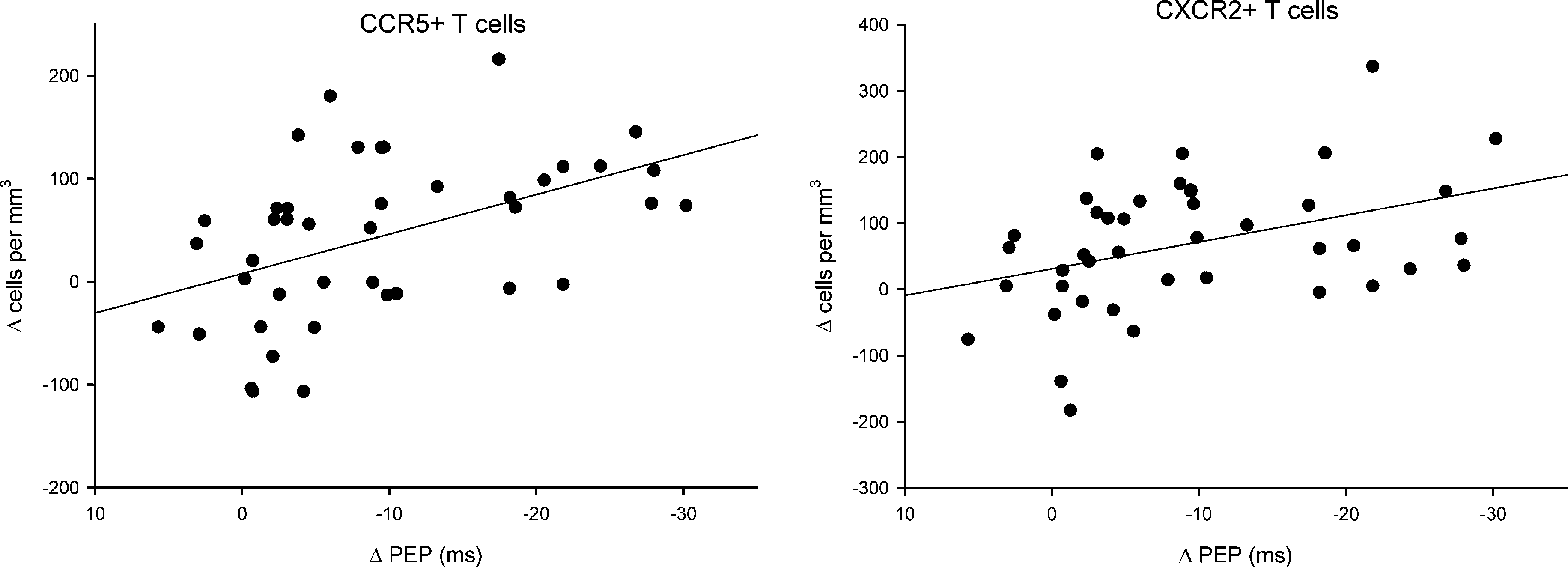 J.A. Bosch et al. / Brain, Behavior, and Immunity 17 (2003) 251–259
Table 4Pearson correlation coefficients and SpearmanÕs rank-order correlation coefficients (between brackets) of the association between PEPreactivity and changes in T cell numbers
Fig. 2. Scatter plots showing the relation between PEP reactivity and mobilization of CCR5+ and CXCR2+ T cells.
J.A. Bosch et al. / Brain, Behavior, and Immunity 17 (2003) 251–259
Table 4Pearson correlation coefficients and SpearmanÕs rank-order correlation coefficients (between brackets) of the association between PEPreactivity and changes in T cell numbers
Fig. 2. Scatter plots showing the relation between PEP reactivity and mobilization of CCR5+ and CXCR2+ T cells.