Tadalafil appartiene alla classe degli inibitori selettivi della fosfodiesterasi di tipo 5, con un profilo farmacocinetico caratterizzato da un’emivita terminale di circa diciotto ore. Dopo somministrazione orale viene assorbito rapidamente e raggiunge concentrazioni plasmatiche massime in due ore. La biotrasformazione avviene principalmente tramite CYP3A4 con formazione di metaboliti inattivi, escreti in prevalenza con le feci. L’elevato legame alle proteine plasmatiche (>90%) assicura una distribuzione stabile. Nei confronti delle altre molecole della stessa classe, cialis compresse italia è noto per la durata prolungata dell’attività farmacologica.
Productstewardship.eu
Official Journal of the European Communities
adapting to technical progress for the 27th time Council Directive 67/548/EEC on the
approximation of laws, regulations and administrative provisions relating to the classification,
packaging and labelling of dangerous substances(*)
THE COMMISSION OF THE EUROPEAN COMMUNITIES,
Having regard to the Treaty establishing the European
The texts in Annexes I and II to this Directive are added to
Part B of Annex V to Directive 67/548/EEC.
Having regard to Council Directive 67/548/EEC of 27 June
1967 on the approximation of laws, regulations and
administrative provisions relating to the classification,
packaging and labelling of dangerous substances (1), as last
amended by European Parliament and Council Directive
Member States shall bring into force the laws, regulations
1999/33/EC (2), and in particular Article 28 thereof,
and administrative provisions necessary to comply with this
Directive by 1 October 2001 at the latest. They shall forthwith
When Member States adopt those provisions, they shall
contain a reference to this Directive or be accompanied by
(1) Annex V to Directive 67/548/EEC lays down the methods
such a reference on the occasion of their official publication.
for the determination of the physico-chemical properties,
Member States shall determine how such reference is to be
toxicity and ecotoxicity of substances and preparations. It
is necessary to adapt that Annex to technical progress.
Member States shall communicate to the Commission the
main provisions of national law which they adopt in the field
(2) According to Article 7(2) of Council Directive
covered by this Directive and a correlation table between this
86/609/EEC of 24 November 1986 on the approximation
Directive and the national provisions adopted.
of laws, regulations and administrative provisions of the
Member States regarding the protection of animals used
for experimental and other scientific purposes (3), an
experiment entailing the use of animals shall not be
performed if another scientifically satisfactory method to
This Directive shall enter into force on the third day following
obtain the result sought is reasonably and practicably
its publication in the Official Journal of the European
(3) The Commission intends to introduce into Annex V to
Directive 67/548/EEC certain alternative testing methods
not entailing the use of animals, in order to make them
This Directive is addressed to the Member States.
available for the testing of chemicals according to Article
(4) The measures provided for in this Directive are in
accordance with the opinion of the Committee on the
Adaptation to Technical Progress of the Directives for the
Elimination of Technical Barriers to Trade in Dangerous
(*) Adopted before the 26th adaptation.
Official Journal of the European Communities
Two in vitro test for skin corrosivity, the rat skin transcutaneous electrical resistance (TER) assay and a test
employing a human skin model, have been endorsed as scientifically valid by the European Centre for the
Validation of Alternative Methods (ECVAM, Joint Research Centre, European Commission) (1) (2) (3). The
ECVAM validation study demonstrated that both tests were able to reliably discriminate between known skin
corrosives and non-corrosives. Furthermore, the test protocol based on a human skin model enabled correct
distinction between degrees of corrosive effects (known severe skin corrosives, R35, and other skin
corrosives, R34) (2). Description and procedures for both tests are given; the choice of which test to use
depends on the specific requirements and preferences of the user.
See also General Introduction, Part B.
Skin corrosion: the production of irreversible tissue damage in the skin following the application of a test
None specified, but see points 1.5.3.4 and 1.7.2.3.
Principle of the test method rat skin TER assay
The test material is applied for up to 24 hours to the epidermal surfaces of skin discs taken from the pelts of
humanely killed young rats. Corrosive materials are identified by their ability to produce a loss of normal
stratum corneum integrity and barrier function, which is measured as a reduction in the inherent TER below
a threshold level (5 kh) (4) (5). Irritant and non-irritant materials do not reduce the TER below the threshold
level. A dye-binding step can be incorporated into the test procedure for surfactants and neutral organics (for
definition see reference (6)) to reduce the number of false positive results obtained specifically with these
Description of the test method rat skin TER assay
Young (20-23 days) rats (Wistar or a comparable strain) are required for the preparation of skin discs. The
dorsal and flank hair is carefully removed with small animal clippers. The animals are then washed by
careful wiping, whilst submerging the area in antibiotic solution (containing, for example, streptomycin,
penicillin, chloramphenicol and amphotericin at concentrations effective in inhibiting bacterial growth).
Animals are washed with antibiotics again on the third or fourth day after the first wash, and are used
within 3 days (animals must not be older than 31 days for pelt preparation).
Animals are humanely killed. The dorsal skin of each animal is then removed and stripped of excess fat by
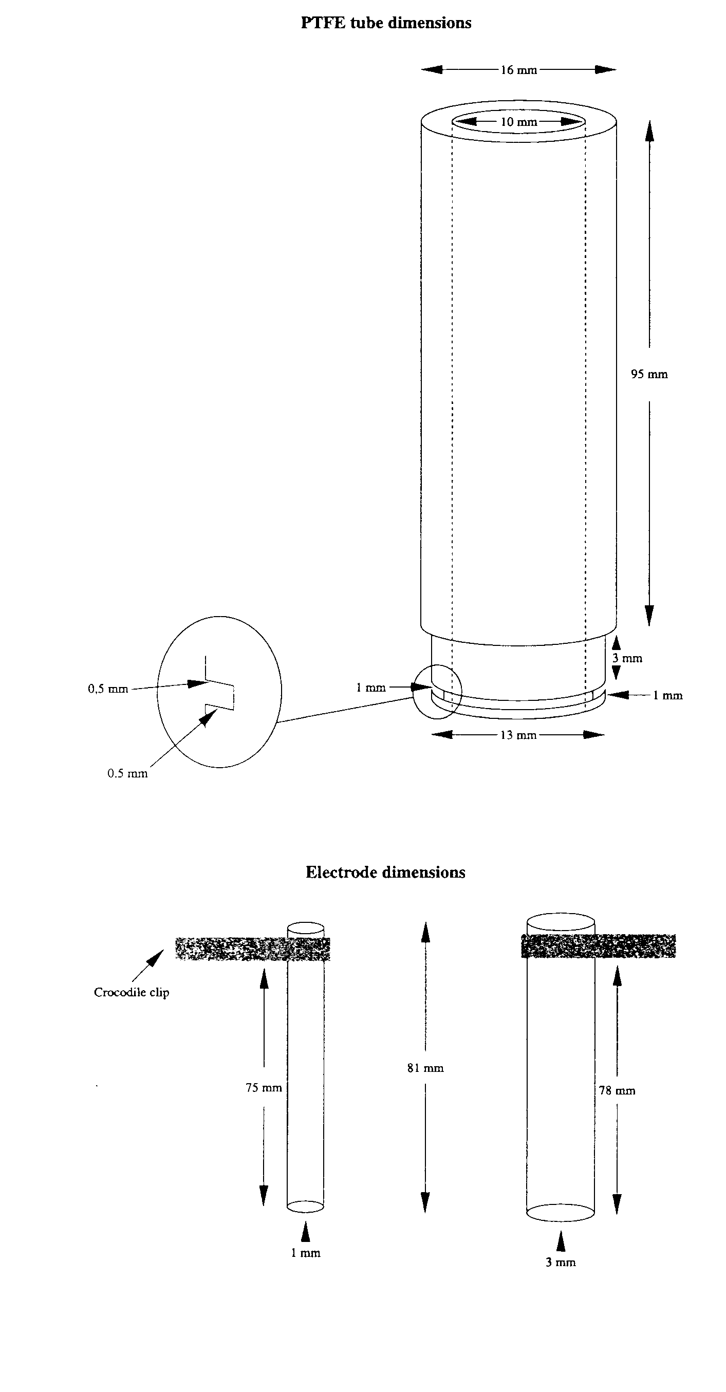
carefully peeling it away from the skin. The pelt is placed over the end of a polytetrafluoroethylene (PTFE)
tube, ensuring that the epidermal surface is in contact with the tube. A rubber O ring is press-fitted over
the end of the tube to hold the skin in place and excess tissue is trimmed away. Tube and O ring
dimensions are shown in Figure 1. The rubber O ring is then carefully sealed to the end of the PTFE tube
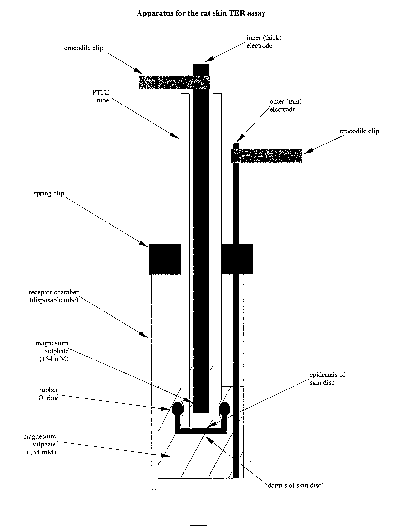
with petroleum jelly. The tube is supported by a spring clip inside a receptor chamber contianing
magnesium sulphate solution (154 mM) (Figure 2).
1.5.3.1. A p p l i c a t i o n o f t h e t e s t m a t e r i a l
Liquid test substances (150 µl) are applied to the epidermal surface inside the tube (Figure 2). When testing
solid materials, a sufficient amount of the solid is applied to the disc to ensure that the whole surface of the
epidermis is covered. Deionised water (150 µl) is then added on top of the solid and the tubes are gently
agitated. Test substances should have maximum contact with the skin. For some solids this may be achieved
by warming up to 30°C to melt the test substance, or by grinding to produce a granular material or powder.
Official Journal of the European Communities
Three skin discs are used for each test substance. Test substances are applied for 24 hours (see also 1.5.3.4).
The test substance is removed by washing with a jet of tap water at up to 30°C until no further material can
be removed. The removal of test substances which have solidified in the tube can be facilitated by jet
washing with warm water at approximately 30°C.
The TER is measured by using a low-voltage, alternating current databridge (e.g. AIM 401 or 6401, or
equivalent). Prior to measuring the electrical resistance, the surface tension of the skin is reduced by adding a
sufficient volume of 70% ethanol to cover the epidermis. After a few seconds the ethanol is removed by
inverting the tube, and the tissue is then hydrated by the addition of 3 ml magnesium sulphate solution (154
mM). The databridge electrodes are placed on either side of the skin disc to take the resistance measurement
in kh/skin disc (Figure 2). Electrode dimensions and the length of the electrode exposed below the crocodile
clips are shown in Figure 1. The inner (thick) electrode clip is rested on the top of the PTFE tube during
resistance measurement, to ensure that a consistent length of electrode is submerged in the magnesium
sulphate solution. The outer (thin) electrode is positioned inside the receptor chamber so that it rests on the
bottom of the chamber. The distance between the bottom of the spring clip and the bottom of the PTFE
tube is maintained as a constant (Figure 1), since this distance affects the resistance value obtained.
Note that if the measured resistance value is greater than 20 kh, this may be due to the test substance
coating the epidermal surface of the sking disc. Removal of this coating can be attempted, for example, by
sealing the PTFE tube with a gloved thumb and shaking it for approximately 10 seconds; the magnesium
sulphate solution is discarded and the resistance measurement is repeated with fresh magnesium sulphate.
The mean TER results are accepted on condition that concurrent positive and negative control values fall
within the acceptable ranges for the method. The suggested control substances and their associated
acceptable resistance ranges for the methodology and apparatus described are:
1.5.3.3. M o d i f i e d p r o c e d u r e f o r s u r f a c t a n t s a n d n e u t r a l o r g a n i c s
If the TER values of test substances which are either surfactants or neutral organics are less than or equal to
5 kh, an assessment of dye penetration can be carried out on the tissues. This procedure will determine
whether the results are false positives (2).
1.5.3.3.1. Sulforhodamine B dye application and removal
Following initial treatment with the test substance, 150 µl of a 10% (w/v) dilution of sulforhodamine B dye
in distilled water is applied to the epidermal surface of each skin disc for 2 hours. The skin discs are then jet
washed with tap water at up to room temperature for approximately 10 seconds to remove any
excess/unbound dye. Each skin disc is carefully removed from the PTFE tube and placed in a vial (e.g. a 20 ml
glass scintillation vial) containing deionised water (8 ml). The vials are agitated gently for 5 minutes to
remove any further excess/unbound dye. This rinsing procedure is then repeated, after which the skin discs
are removed and placed into vials containing 5 ml of 30% (w/v) sodium dodecyl sulphate (SDS) in distilled
water and are incubated overnight at 60°C. After incubation, each skin disc is removed and discarded and
the remaining solution is centrifuged for 8 minutes at 21°C (relative centrifugal force p 175). A 1 ml
sample of the supernatant is then diluted 1 in 5 (v/v) (i.e. 1 ml + 4 ml) with 30% (w/v) SDS in distilled
water. The optical density (OD) of the solution is measured at approximately 565 nm.
The sulforhodamine B dye content per disc is calculated from the OD values (sulforhodamine B dye molar
extinction coefficient at 565 nm = 8,7×10⁴; molecular weight = 580). The sulforhodamine B dye content is
determined for each skin disc and a mean dye content is then calculated for the replicates. The mean dye
binding results are accepted on condition that concurrent control values fall within the acceptable ranges for
the method. Suggested acceptable dye content ranges for the control substances for the methodology and
Official Journal of the European Communities
1.5.3.4. A d d i t i o n a l i n f o r m a t i o n
Test substances can also be applied to the skin discs for shorter periods (e.g. 2 hours) to identify those
materials which are severely corrosive. However, in the validation study, the TER assay was found to
overestimate the corrosive potential of several test chemicals following their application to the skin discs for
2 hours (2), although it enabled the correct identification of corrosives and non-corrosives after a 24-hour
The properties and dimensions of the test apparatus and the experimental procedure used may influence the
TER values obtained. The 5 kh corrosive threshold was developed from data obtained with the specific
apparatus and procedure described in this method. Different threshold and control values may apply if the
test conditions are altered significantly. Therefore, it is recommended that the methodology and resistance
threshold value are calibrated by testing a series of reference standards chosen from the chemicals used in
Principle of the test method human skin model assay
The test material is applied topically for up to 4 hours to a three-dimensional human skin model,
comprising a reconstructed epidermis with a functional stratum corneum. Corrosive materials are identified
by their ability to produce a decrease in cell viability (as determined, for example, by using the MTT
reduction assay) below defined threshold levels at specified exposure periods. The principle of the essay is in
accordance with the hypothesis that chemicals which are corrosive are those which are able to penetrate the
stratum corneum (by diffusion or erosion) and are sufficiently cytotoxic to cause cell death in the underlying
Description of the test method human skin model assay
Human skin models can come from various sources, but they must meet certain criteria. The model must
have a functional stratum corneum with an underlying layer of living cells. The barrier function of the
stratum corneum must be adequate. This can be shown by demonstrating the model's resistance to
cytotoxicity following the application of substances which are known to be cytotoxic to cells, but which do
not normally pass through the stratum corneum. The model must be shown to give reproducible results
under defined experimental conditions.
The viability of the living cells in the model must be sufficiently high to discriminate well between the
positive and negative control substances. Cell viability (for example, as measured by the amount of MTT
reduction, i.e. an OD value) following exposure to the negative control substance must fall within acceptable
limits for the particular model. Similarly, cell viability values with the positive control substance (relative to
those for the negative control) must fall within specified limits. Most importantly, the prediction model used
must have been shown to meet the international validation standard (2).
1.7.2.1. A p p l i c a t i o n o f t h e t e s t m a t e r i a l
For liquid materials, sufficient test substance must be applied to cover the skin surface (a minimum of
25 µl/cm2). For solid materials, sufficient test substance must be applied to cover the skin, and it should then
be moistened to ensure good contact with the skin; where appropriate, solids should be ground to a powder
before application. The application method must be shown to be adequate for a wide range of chemical
types (2). At the end of the exposure period, the test material must be carefully washed from the skin surface
1.7.2.2. C e l l v i a b i l i t y m e a s u r e m e n t s
Any quantitative, validated, method can be used to measure cell viability. The most frequently used assay is
MTT reduction, which has been shown to give accurate and reproducible results in various laboratories (2).
The skin disc is placed in an MTT solution of 0,3 mg/ml at 20-28°C for 3 hours. The precipitated blue
formazan product is then extracted (solvent extraction) and the concentration of the formazan is measured
by determining the OD at a wavelenght between 545 and 595 nm.
Official Journal of the European Communities
1.7.2.3. A d d i t i o n a l i n f o r m a t i o n
The skin model used, and the exact protocol of exposure time and washing procedures, etc. will have a
major impact on the cell viability results. It is recommended that the methodology and prediction model are
calibrated by testing a series of reference standards chosen from the chemicals used in the ECVAM validation
study (3). It is critical that the method used has been shown to be reproducible within and between
laboratories for a wide range of chemicals, in accordance with international standards. As a minimum, the
method should meet the criteria for scientific validity defined previously (2), and the results of such a
validation study must be published in a peer-reviewed scientific journal.
Resistance values (kh) for the test material, positive and negative controls, and any standard reference
chemicals should be reported in tabular form, including data for replicates/repeat experiments, mean values
OD values and calculated percentage cell viability data for the test material, positive and negative controls,
and any standard reference chemicals should be reported in tabular form, including data for replicates/repeat
experiments, mean values and the classification derived.
If the mean TER value obtained for the test substance is greater than 5 kh, then it is non-corrosive. If the
TER value is less than or equal to 5 kh, and the test substance is not a surfactant or neutral organic, then it
For surfactants or neutral organics which give TER values less than or equal to 5 kh, dye penetration can be
carried out. If the mean disc dye content is greater than or equal to the mean disc dye content of the 36%
HCl positive control obtained concurrently, then the test substance is a true positive and is therefore
corrosive. If the mean disc dye content is less than the mean disc content of the 36% HCl positive control
obtained concurrently, then the test substance is a false positive and is therefore non-corrosive.
The negative control OD value represents 100% cell viability; hence, the OD values obtained for each test
sample can be used to calculate a percentage viability relative to the negative control. The cut-off percentage
cell viability value distinguishing corrosive from non-corrosive test materials (or discriminating between
different corrosive classes) must be clearly defined in the prediction model before the method is validated,
and the subsequent validation study must show that the cut-off value is appropriate (2).
The test report must include at least the following information:
identification data, physical nature and, where relevant, physicochemical properties. Similar information
should be provided for reference substances, if used.
description and justification of any modifications.
Official Journal of the European Communities
Results: tabulation of resistance values (TER assay) or percentage cell viability values (human skin model assay)
for the test material, positive and negative controls and any standard reference chemicals, including data
for replicates/repeat experiments and mean values,
description of any other effects observed.
REFERENCES(1) ECVAM (1998), ECVAM News & Views, ATLA 26, pp. 275-280. (2) Fentem, J.H., Archer, G.E.B., Balls, M., Botham, P.A., Curren, R.D., Earl, L.K., Esdaile, D.J., Holzhutter,
H-G. & Liebsch, M. (1998), The ECVAM international validation study on in vitro tests for skin
corrosivity. 2. Results and evaluation by the Management Team, Toxicology in Vitro 12, pp. 483-524.
(3) Barratt, M.D., Brantom, P.G., Fentem, J.H., Gerner, I., Walker, A.P. & Worth, A.P. (1998), The ECVAM
international validation study on in vitro tests for skin corrosivity. 1. Selection and distribution of the test
chemicals, Toxicology in Vitro 12, pp. 471-482.
(4) Oliver, G.J.A., Pemberton, M.A. & Rhodes, C. (1986), An in vitro skin corrosivity test modifications
and validation, Food & Chemical Toxicology 24, pp. 507-512.
(5) Botham, P.A., Hall, T.J., Dennett, R., McCall, J.C., Basketter, D.A., Whittle, E., Cheeseman, M., Esdaile,
D.J. & Gardner, J. (1992), The skin corrosivity test in vitro: results of an interlaboratory trial, Toxicology in
(6) Worth, A.P., Fentem, J.H., Balls, M., Botham, P.A., Curren, R.D., Earl, L.K., Esdaile, D.J. & Liebsch, M.
(1998), An evaluation of the proposed OECD testing strategy for skin corrosion, ATLA 26, pp.
(7) Botham, P.A., Chamberlain, M., Barratt, M.D., Curren, R.D., Esdaile, D.J., Gardner, J.R., Gordon, V.C.,
Hildebrand, B., Lewis, R.W., Liebsch, M., Logemann, P., Osborne, R., Ponec, M., Regnier, J.F., Steiling,
W., Walker, A.P. & Balls, M. (1995), A prevalidation study on in vitro skin corrosivity testing. The report
and recommendations of ECVAM workshop 6, ATLA 23, pp. 219-255.
Official Journal of the European Communities
Official Journal of the European Communities
Official Journal of the European Communities
B.41. PHOTOTOXICITY IN VITRO 3T3 NRU PHOTOTOXICITY TEST
Phototoxicity is defined as a toxic response that is elicited after the first exposure of skin to certain chemicals
and subsequent exposure to light, or that is induced similarly by skin irradiation after systemic administration
Information derived from the in vitro 3T3 NRU phototoxicity test serves to identify the phototoxic potential
of a test substance, i.e. the existence or absence of possible hazards likely to arise from a test substance in
association with exposure to UV and visible light.
Since the toxicological endpoint of the in vitro test is determination of photocytotoxicity, induced by the
combined action of a chemical and light, compounds that are phototoxic in vivo after systemic application
and distribution to the skin, as well as compounds that act as photoirritants after topical application to the
skin, can be identified by the test.
The in vitro 3T3 NRU phototoxicity test was developed and validated in a joint EU/COLIPA project from
1992-1997 (1) (2) (3), to establish a valid in vitro alternative to the various in vivo tests in use. In 1996 an
OECD workshop recommended an in vitro tier testing approach for phototoxicity assessment (4).
Results from the in vitro 3T3 NRU phototoxicity test were compared with acute phototoxicity/photoirritation
effects in vivo in animals and humans, and the test has been shown to give excellent predictivity for these
effects. The test is not designed to predict other adverse effects that may arise from the combined action of a
chemical and light, e.g. photogenotoxicity, photoallergy, and photocarcinogenicity, although many chemicals
which show these specific properties will react positive in the in vitro 3T3 NRU phototoxicity test. In
addition, the test is not designed to permit an assessment of phototoxic potency.
A sequential approach to phototoxicity testing of chemicals is set out in the Appendix.
Irradiance: the intensity of ultraviolet (UV) or visible light incident on a surface, measured in W/m2 or
Dose of light: the quantity (= intensity×time) of ultraviolet (UV) or visible radiation incident on a surface,
expressed in Joules (= W×s) per surface area, e.g. J/m2 or J/cm2.
UV light wavebands: the designations recommended by the CIE (Commission Internationale de L'Eclairage) are:
UVA (315-400 nm), UVB (280-315 nm) and UVC (100-280 nm). Other designations are also used: the
division between UVB and UVA is often placed at 320 nm, and the UVA may be divided into UV-A1 and
UV-A2 with a division made at about 340 nm.
Cell viability: parameter measuring total activity of a cell population (e.g. uptake of the vital dye neutral red
into cellular lysosomes) which, depending on the endpoint measured and the test design used, correlates with
the total number and /or vitality of the cells.
Relative cell viability: cell viability expressed in relation to negative (solvent) controls which have been taken
through the whole test procedure (either +UV or ¯UV), but not treated with a test chemical.
Prediction model: an algorithm used to transform the results of a toxicity test into a prediction of toxic
potential. In the present test guideline, PIF and MPE can be used for transformation of the results of the in
vitro 3T3 NRU phototoxicity test into a prediction of phototoxic potential.
PIF (photo irritation factor): a factor generated by comparing two equally effective cytotoxic concentrations
(EC50) of the test chemical obtained in the absence (¯UV) and in the presence (+UV) of a non-cytotoxic
MPE (mean photo effect): a novel measure derived from mathematical analysis of the complete shape of two
concentration response curves obtained in the absence (¯UV) and in the presence (+UV) of a noncytotoxic
Official Journal of the European Communities
Phototoxicity: an acute toxic response that is elicited after the first exposure of skin to certain chemicals and
subsequent exposure to light, or that is induced similarly by skin irradiation after the systemic administration
Photoirritation: a sub-species of the term phototoxicity, which is used to describe only those phototoxic
reactions which are produced at the skin after exposure to chemicals (topically or orally). These photoxic
reactions lead always to non-specific cell damage (sunburn like reactions).
Photoallergy: an acquired immunological reactivity, which does not occur on first treatment with chemical and
light, and needs an induction period of one or two weeks before skin reactivity can be demonstrated.
Photogenotoxicity: a genotoxic response observed with a genetic endpoint, which is elicited after the exposure
of cells to a non-genotoxic dose of UV/visible light and a non-genotoxic chemical.
Photocarcinogenicity: carcinogenicity induced by repeated application of light and a chemical. The term photo
co-carcinogenesis, is used if UV induced tumorigenesis is enhanced by a chemical.
Besides the positive control chemical chlorpromazine which should be concurrently tested in each assay, for
newly establishing the 3T3 NRU phototoxicity test it is recommended to use as reference chemicals a subset
from the chemicals used in interlaboratory trials with the present test (1) (3) (13).
Many types of chemicals have been reported to induce phototoxic effects (5) (6) (7) (8). The only common
feature is their ability to absorb light energy within the sunlight region. According to the first law of
photochemistry (Grotthaus-Draper's Law) photoreaction requires sufficient absorption of light quanta. Thus,
before biological testing according to the present test guideline is considered, a UV/vis absorption spectrum
of the test chemical should be determined (e.g. according to OECD Test Guideline 101). If the molar
extinction /absorption coefficient is less than 10 litre×mol¯1×cm¯1, the chemical has no photoreactive
potential and does not need to be tested in the in vitro 3T3 NRU phototoxicity test or any other biological
test for adverse photochemical effects (Appendix).
Four mechanisms have been identified by which absorption of light by a (chemical) chromophore can result
in a phototoxic response (7). All of them result in cell damage. Therefore, the in vitro 3T3 NRU phototoxicity
test is based on a comparison of the cytotoxicity of a chemical when tested in the presence and in the
absence of exposure to a non-cytotoxic dose of UVA/vis light. Cytotoxicity in this test is expressed as a
concentration dependent reduction of the uptake of the vital dye neutral red (NR) (9) 24 hours after
treatment with the test chemical and irradiation.
Balb/c 3T3 cells are maintained in culture for 24 h for the formation of monolayers. Two 96-well plates per
test chemical are then preincubated with eight different concentrations of the chemical for 1 h. Thereafter
one of the two plates is exposed to a non-cytotoxic UVA/vis light dose of 5 J/cm2 UVA ( +UV experiment),
whereas the other plate is kept in the dark (¯UV experiment). In both plates, the treatment medium is then
replaced by culture medium and after another 24 h of incubation, cell viability is determined by neutral red
uptake (NRU) for 3 h. Relative cell viability, expressed as percentage of untreated negative controls, is
calculated for each of the eight test concentrations. To predict the phototoxic potential, the concentration
responses obtained in the presence (+UV) and in the absence (¯UV) of irradiation are compared, usually at
the EC50 level, i.e. at the concentration inhibiting cell viability by 50% cf. untreated controls.
UVA sensitivity of the cells, historical data: cells should be regularly checked for sensitivity to UVA. Cells are
seeded at the density used in the in vitro 3T3 NRU phototoxicity test, irradiated the next day with UVA doses
from 1-9 J/cm2, and cell viability is determined one day later using the NRU assay. Cells meet the quality
criteria, if their viability after irradiation with 5 J/cm2 UVA is not less than 80% of the viability of dark
controls. At the highest UVA dose of 9 J/cm2, viability should not be less than 50% of that of dark controls.
This check should be repeated about every 10th passage of the cells.
UVA sensitivity of the negative control cells, current test: the test meets the quality criteria if negative controls
(cells in Earl's balanced salt solution (EBSS) with or without 1% dimethylsulfoxide (DMSO) or 1% ethanol
(EtOH)) in the +UVA experiment show a viability of not less than 80% of that of non-irradiated cells in the
same solvent of the concurrent dark experiment (¯UVA).
Official Journal of the European Communities
Viability of negative controls: the absolute optical density (OD540 NRU) measured in the NR extract of the
negative controls indicates whether the 1×10⁴ cells seeded per well have grown with normal doubling time
during the two days of the assay. A test meets the acceptance criteria if the mean OD540 NRU of untreated
Positive control: a known phototoxic chemical shall be tested concurrently with each in vitro 3T3 NRU
phototoxicity test. Chlorpromazine (CPZ) was used as positive control in the EU/COLIPA validation study and
is therefore recommended. For CPZ tested with the standard protocol in the in vitro 3T3 NRU phototoxicity
test, the following test acceptance criteria were defined: CPZ irradiated (+UVA): EC50 = 0,1 to 2,0 µg/ml, CPZ
non-irradiated (¯UVA): EC50 = 7,0 to 90,0 µg/ml. The photo irritation factor (PIF), i.e. the shift of EC50
Other known phototoxic chemicals, suitable for the chemical class or solubility characteristics of the test
chemical being evaluated, may be used as the concurrent positive controls, in place of CPZ. In this case,
based on historical data, the ranges of EC50 values and PIF or MPE (mean photo effect) should be adequately
defined as acceptance criteria for the test.
A permanent mouse fibroblast cell line Balb/c 3T3, clone 31 either from ATCC or from ECACC was
used in the validation study, and is therefore recommended. Other cells or cell lines may be successfully used
with the same test protocol, if the culture conditions are adapted to the specific needs of the cells, but
Cells should be checked regularly for the absence of mycoplasma contamination and should only be used if
the results of such checking was satisfactory.
Since the UVA sensitivity of cells may increase with the number of passages, Balb/c 3T3 cells of the lowest
obtainable passage number should be used, preferably less than 100. It is important that UVA sensitivity of
the Balb/c 3T3 cells is regularly checked according to the quality control procedure described in this
1.7.1.2. M e d i a a n d c u l t u r e c o n d i t i o n s
Appropriate culture media and incubation conditions should be used for routine cell passage and during the
test procedure. For Balb/c 3T3 cells, these are DMEM supplemented with 10% new-born calf serum, 4 mM
glutamine, penicillin and streptomycin; and humidified incubation at 37°C /7,5% CO2. It is particularly
important that cell culture conditions ensure a cell cycle time within the normal historical range of the cells
1.7.1.3. P r e p a r a t i o n o f c u l t u r e s
Cells from frozen stock cultures are seeded in culture medium at an appropriate density and subcultured at
least once before they are used in the in vitro 3T3 NRU phototoxicity test.
For the phototoxicity test cells are seeded in culture medium at a density such that cultures will not reach
confluence by the end of the test, i.e. when cell viability is determined 48 h after the seeding of the cells. For
Balb/c 3T3 cells grown in 96-well plates, 1×10⁴ cells per well is the recommended cell density.
For each test chemical, cells are seeded identically in two separate 96-well plates, which are then taken
concurrently through the whole test procedure under identical culture conditions, except for the time period
where one of the plates is irradiated (+UVA/vis) and the other one is kept in the dark (¯UVA/vis).
1.7.1.4. M e t a b o l i c a c t i v a t i o n
Whereas the use of metabolising systems is a general requirement for all in vitro tests for the prediction of
genotoxic and carcinogenic potential, up to now, in the case of phototoxicology, no chemical is known for
which metabolic transformation is needed for the chemical to act as a phototoxin in vivo or in vitro. Thus, it
is neither considered necessary nor scientifically justified for the present test to be performed with a
Official Journal of the European Communities
1.7.1.5. T e s t c h e m i c a l / p r e p a r a t i o n
Test chemicals must be freshly prepared immediately prior to use, unless stability data demonstrate the
acceptability of storage. Preparation under red light may be required when rapid photodegradation is likely to
Test chemicals should be dissolved in buffered salt solutions, e.g. Earl's balanced salt solution, (EBSS) or
phosphate buffered saline (PBS), which, to avoid interference during irradiation, must be free from protein
components and light absorbing pH indicator colours.
Test chemicals of limited solubility in water should be dissolved in appropriate solvents at 100-fold the
desired final concentration and then diluted 1:100 with the buffered salt solution. If a solvent is used it must
be present at a constant volume of 1% (v/v) in all cultures, i.e. in the negative controls as well as in all
concentrations of the test chemical.
Dimethylsulphoxide (DMSO) and ethanol (EtOH) are the recommended solvents. Other solvents of low
cytotoxicity (e.g. acetone) may be appropriate, but they should carefully be assessed for specific properties,
e.g. reaction with the test chemical, quenching of the phototoxic effect, radical catching properties.
Vortex mixing and/or sonication and/or warming to 37°C may be used, if necessary, to aid solubilisation.
1.7.1.6. U V i r r a d i a t i o n / p r e p a r a t i o n
Light source: the choice of an appropriate light source and appropriate filtering is the most crucial factor in
phototoxicity testing. UVA and visible regions are usually associated with photosensitisation (7) (10), whereas
UVB is of less relevance and is directly highly cytotoxic, increasing its cytotoxicity through 1000 fold from
313 to 280 nm (11). Criteria for the choice of an appropriate light source should include the essential
requirement that the light source emits wavelengths absorbed by the test chemical and that the dose of light
(achievable in a reasonable time) should be sufficient for the detection of known photosensitisers.
Furthermore, the wavelengths and doses employed should not be unduly deleterious to the test system, which
includes the emission of heat (infra-red region).
The simulation of sunlight with solar simulators is considered the optimal light source. Both, Xenon arcs and
(doped) mercury-metal halide arcs are used in solar simulators. The latter have the advantage of emitting less
heat and of being cheaper, but the match to sunlight is not perfect. Since all solar simulators emit significant
quantities of UVB, they should be suitably filtered to attenuate the highly cytotoxic UVB wavelengths.
For the in vitro 3T3 NRU phototoxicity test an irradiance spectrum practically devoid of UVB should be used
(UVA:UVB p 1:20). An example of the spectral irradiance distribution of the filtered solar simulator used in
the validation study of the in vitro 3T3 NRU phototoxicity test has been published (3).
Dosimetry: the intensity of light (irradiance) should be regularly checked before each phototoxicity test, by
using a suitable broadband UV-meter. The UV-meter must have been calibrated to the source. The
performance of the UV-meter should be checked, and for this purpose, the use of a second, reference
UV-meter of the same type and identical calibration is recommended. Ideally, at greater intervals, a
spectroradiometer should be used to measure the spectral irradiance of the filtered light source and to check
the calibration of the broadband UV-meter, but such instruments require skilled operation by appropriately
A dose of 5 J/cm2 (UVA) was determined in the validation study to be non-cytotoxic to Balb/c 3T3 cells and
sufficiently potent to excite even weak phototoxic chemicals. To achieve 5 J/cm2 within a time period of 50
min, irradiance has to be adjusted to 1,666 mW/cm2. If another cell line or a different light source are used,
the UVA dose may have to be slightly adapted, by using the criteria of being non-deleterious to the cells and
sufficient to detect standard phototoxins. The time of light exposure is calculated in the following way:
The maximum concentration of a test chemical should not exceed 100 µg/ml, since all phototoxic chemicals
were detected at lower concentrations, whereas at higher concentrations the incidence of false positives
(overpredictions) increases (13). The pH of the highest concentration of the test chemical should be
Official Journal of the European Communities
The ranges of concentrations of a chemical tested in the presence (+UVA) and in the absence (¯UVA) of light
should be adequately determined in preceding range-finder experiments. Range and intercept of a
concentration series shall be adjusted in such a way that concentration-response curves are sufficiently
supported by experimental data. Geometric concentration series (with a constant dilution factor) should be
Prepare a cell suspension of 1 × 1055 cells/ml in culture medium and dispense 100 µl culture medium only
into the peripheral wells of a 96-well tissue culture microtiter plate (= blanks). In the remaining wells,
dispense 100 µl of a cell suspension of 1 × 10⁵ cells/ml (= 1 × 10⁴ cells/well). For each test chemical, prepare
two plates: one for determination of cytotoxicity (¯UVA), and the other for determination of
Incubate the cells for 24 h (7,5% CO2, 37°C) until they form a half-confluent monolayer. This incubation
period allows for cell recovery and adherence, and for exponential growth.
After incubation, decant the culture medium from the cells and wash twice with 150 µl EBSS/PBS per well.
Add 100 µl of EBSS/PBS containing the appropriate concentration of test chemical or just solvent (negative
control). Apply 8 different concentrations of the test chemical. Incubate cells with the test chemical in the
dark for 60 minutes (7,5% CO2, 37°C).
To perform the (+UVA) part of the assay, irradiate the cells at room temperature for 50 minutes through the
lid of the 96-well plate with 1,7 mW/cm2 UVA (= 5 J/cm2). Ventilate with a fan to prevent H2O
condensation under the lid. Keep duplicate plates (¯UVA) at room temperature in a dark box for 50 min
Decant test solution and wash twice with 150 µl EBSS/PBS. Replace EBSS/PBS with culture medium and
incubate (7,5% CO2, 37°C) overnight (18-22 h).
Examine the cells under a phase-contrast microscope. Record changes in morphology of the cells due to
cytotoxic effects of the test chemical. This check is recommended, to exclude experimental errors, but these
records are not used for evaluation of cytotoxicity or phototoxicity
Wash the cells with 150 µl prewarmed EBSS/PBS. Remove the washing solution by gentle tapping. Add
100 µl NR medium and incubate at 37°C, in a humidified atmosphere of 7,5% CO2, for 3 h.
After incubation, remove the NR medium, and wash the cells with 150 µl EBSS/PBS. Decant and blot
EBSS/PBS totally. (Optionally: centrifuge reversed plate.)
Add exactly 150 µl NR desorb solution (freshly prepared ethanol/acetic acid)
Shake microtiter plate rapidly on a microtiter plate shaker for 10 min, until the NR has been extracted from
the cells and has formed a homogeneous solution.
Measure the optical density of NR extract at 540 nm in a spectrophotometer, using blanks as a reference.
Save the data in appropriate file format (e.g. ASCII) for subsequent analysis.
(1) Additional details can be found in reference 12.
Official Journal of the European Communities
The data should permit a meaningful analysis of the concentration-response obtained in the presence and in
the absence of UVA/vis irradiation. If cytotoxicity is found, both the concentration range and the intercept of
individual concentrations should be set in such a way as to allow the fit of a curve to experimental data. Due
to the fact that a test chemical might not be cytotoxic up to the defined limit concentration of 100 µg/ml in
the dark experiment (¯UVA), but highly cytotoxic when irradiated (+UVA), the concentration ranges to be
tested in both parts of the experiment may need to differ by orders of magnitude to fulfil the requirement of
adequate data quality. If no cytotoxicity is found in both parts of the experiment (¯UVA and +UVA), testing
with a great intercept between single doses up to the highest concentration is sufficient.
There is no requirement for verification of a clear positive result by performing a repeat experiment. In
addition, clear negative results need not to be verified, provided the test chemical was tested at sufficiently
high concentrations. In such cases, one main experiment, supported by one or more range-finding
preliminary experiments, is sufficient.
Tests with borderline results near to the cut-off line of the prediction model should be repeated for
If repeat testing is considered necessary, then variation of the experimental conditions may be important to
achieve a clear result. A key variable in this test is preparation of solutions of the test chemical. Hence,
variation of these conditions (co-solvent, trituration, sonication) may be most relevant in the repetition of a
test. Alternatively, variation of the pre-irradiation incubation time may be considered. A shorter time can be
relevant for water-unstable chemicals.
Where possible, the concentration of a test chemical reflecting a 50% inhibition of the cellular NRU (EC50) is
determined. This can be done by applying any appropriate non-linear regression procedure (preferably a Hill
function or logistic regression) to the concentration-response data, or by using other fitting procedures (14).
Before using an EC50 for further calculations, the quality of the fit should be appropriately checked.
Alternatively, graphical fitting methods can be used to calculate the EC50. In this case, the use of probability
paper is recommended (x-scale: log, y-scale: probit), as in many cases the concentration response function
will become almost linear after this transformation.
Evaluation of results (prediction models)
Prediction model version 1: photo-irritation-factor (PIF)
If both, in the presence (+UVA) and in the absence (¯UVA) of light, complete concentration response curves
are obtained, a photo-irritation-factor (PIF) is calculated by means of the following formula:
A PIF < 5, predicts no phototoxic potential, whereas a PIF Å 5 predicts phototoxic potential.
If a chemical is only cytotoxic +UVA and is not cytotoxic when tested ¯UVA, the PIF cannot be calculated,
although this is a result that indicates phototoxic potential. In such cases, a > PIF can be calculated if the
(¯UV) cytotoxicity test is performed up to the highest test concentration (Cmax) and this value is used for
If only a > PIF can be obtained, then any value > 1 predicts phototoxic potential.
If both EC50 (¯UV) and EC50 (+UV) cannot be calculated due to the fact that a chemical does not show any
cytotoxicity up to the highest test concentration, this indicates no phototoxic potential. In such cases, a
formal PIF = *1 is used to characterise the result;
Official Journal of the European Communities
If only a PIF = *1 can be obtained, this predicts no phototoxic potential.
In cases (b) and (c), concentrations achieved in the in vitro 3T3 NRU phototoxicity test should be carefully
taken into consideration when predicting phototoxic potential.
Prediction model version 2: mean-photo effect (MPE)
Alternatively, a novel version of the model for predicting phototoxic potential can be applied, which has
been developed by using data of the EU/COLIPA validation study (15) and tested under blind conditions in a
subsequent study on the in vitro phototoxicity of UV filter chemicals (13). This model overcomes the
limitation of the PIF model in cases where an EC50 cannot be obtained. The model uses the mean photo
effect (MPE), a measure which is based on comparison of the complete concentration response curves. For
application of the MPE model, a special computer software was developed at the Humboldt University
(Berlin), which can be obtained free of charge.
A positive result in the in vitro 3T3 NRU phototoxicity test (PIF Å 5 or MPE Å 0,1) indicates that the test
substance has phototoxic potential. If this result is obtained at concentrations below 10 µg/ml, the test
chemical is also likely to act as phototoxin also under various exposure conditions in vivo. If a positive result
is obtained only at the highest test concentration of 100 µg/ml, further considerations may be necessary for
the assessment of hazard or phototoxic potency. These may include data on penetration, absorption and
possible accumulation of the chemical in the skin, or testing of the chemical in a confirmatory alternative
test, e.g. using a human in vitro skin model.
A negative result from the in vitro 3T3 NRU phototoxicity test (PIF < 5 or MPE < 0,1) indicates that the test
substance was not phototoxic to the cultured mammalian cells under the conditions used. In cases where the
chemical could be tested up to the highest concentration of 100 µg/ml, a negative result indicates that the
chemical has no phototoxic potential, and phototoxicity in vivo may be considered unlikely. In cases where
identical concentration-toxicity responses (EC50 +UV and EC50¯UV) were obtained at lower concentrations,
the interpretation of data would be the same. In contrast, if no toxicity was demonstrated (+UV and ¯UV)
and if aqueous solubility limited concentrations to values less than 100 µg/ml, then compatibility of the test
substance with the assay may be questioned and confirmatory testing should be considered (e.g. using an in
vitro skin model, or an ex vivo skin model or an in vivo test).
The test report must include the following information:
Test chemical: identification data and CAS No, if known, physical nature and purity, physicochemical properties relevant to conduct of the study, stability and photostability, if known.
Solvent: justification for choice of solvent, solubility of the test chemical in this solvent, percentage of solvent present in treatment medium (EBSS or PBS).
Cells: type and source of cells, absence of mycoplasma, number of cell passages, if known, UVA sensitivity of cells, determined with the irradiation equipment, used in the in vitro 3T3 NRU
Test conditions (a) incubation before and after treatment: type and composition of culture medium, incubation conditions (CO2 concentration, temperature, humidity), duration of incubation (pre-treatment, post-treatment).
Official Journal of the European Communities
Test conditions (b) treatment with the chemical:
rationale for selection of concentrations of the test chemical used both in the presence and in the
in case of limited solubility of the test chemical and absence of cytotoxicity, rationale for the highest
type and composition of treatment medium (buffered salt solution),
duration of the chemical treatment.
rationale for selection of the light source used,
spectral irradiance characteristics of the light source,
transmission/absorption characteristics of the filter(s) used,
characteristics of the radiometer and details on its calibration,
distance of the light source from the test system,
UVA irradiance at this distance, expressed in mW/cm2,
duration of the UV/vis light exposure,
UVA dose (irradiance × time), expressed in J/cm2,
temperature employed to cell cultures during irradiation, and for cell cultures concurrently kept in the
incubation conditions (CO2 concentration, temperature, humidity), NR extraction conditions (extractant, duration),
wavelength used for spectrophotometric reading of NR optical density,
second wavelength (reference), if used,
content spectrophotometer blank, if used.
cell viability obtained at each concentration of the test chemical, expressed in percent mean viability of
concentration-response curves, (test chemical concentration v. relative cell viability), obtained in
data analysis of the concentration response curves: if possible, computation/calculation of EC50 (+UVA)
comparison of the two concentration response curves, obtained in the presence and in the absence of
UVA/vis irradiation, either by calculation of the photo irritation factor (PIF), or by calculation of the
classification of phototoxic potential,
test acceptance criteria (a) concurrent negative control:
absolute viability (optical density of NR extract) of irradiated and non irradiated cells, historical data of negative control, mean and standard deviation.
test acceptance criteria (b) concurrent positive control:
EC50 (+UVA) and EC50 (¯UVA) and PIF of positive control chemical, historical data of positive control chemical: EC50 (+UVA) and EC50 (¯UVA) and PIF, mean and
Official Journal of the European Communities
(1) Spielmann, H., Balls, M., Döring, B., Holzhütter, H.G., Kalweit, S., Klecak, G., L'Eplattenier, H., Liebsch,
M., Lovell, W.W., Maurer, T., Moldenhauer, F., Moore, L., Pape, W., Pfannbecker, U., Potthast, J., De
Silva, O., Steiling, W. and Willshaw, A. ( 1994), EEC/COLIPA project on in vitro phototoxicity testing:
First results obtained with a Balb/c 3T3 cell phototoxicity assay, Toxicology in Vitro 8, pp. 793-796.
(2) Anon (1998), Statement on the scientific validity of the 3T3 NRU PT test (an in vitro test for
phototoxicity), European Commission, Joint Research Centre: ECVAM and DGXI/E/2, 3 November
(3) Spielmann, H., Balls, M., Dupuis, J., Pape, W. J. W., Pechovitch, G., De Silva, O., Holzhütter, H. G.,
Clothier, R., Desolle, P., Gerberick, F., Liebsch, M., Lovell, W. W., Maurer, T., Pfannenbecker, U.,
Potthast, J. M., Csato, M., Sladowski, D., Steiling, W. and Brantom, P. (1998), EU/COLIPA In vitro
phototoxicity validation study, results of phase II (blind trial), part 1: the 3T3 NRU phototoxicity test,
Toxicology in Vitro 12, pp. 305-327.
(4) OECD Test Guidelines Programme, ENV/MC/CHEM/TG(96)9: Final Report of the OECD Workshop on
Harmonisation of Validation and Acceptance Criteria of Alternative Toxicological Test Methods, OECD
(5) Lovell, W.W. (1993), A scheme for in vitro screening of substances for photoallergenic potential,
(6) Santamaria, L. and Prino, G. (1972), List of the photodynamic substances, Research progress in organic,
biological and medicinal chemistry Vol. 3 Part 1, North Holland Publishing Co, Amsterdam, pp. XI-XXXV.
(7) Spielmann, H., Lovell, W.W., Hölzle, E., Johnson, B.E., Maurer, T., Miranda, M.A., Pape, W.J.W., Sapora,
O. and Sladowski, D. (1994), In vitro phototoxicity testing: The report and recommendations of ECVAM
(8) Spikes, J.D. (1989), Photosensitization, The science of photobiology, edited by KC Smith, Plenum Press,
(9) Borenfreund, E. and Puerner, J.A. (1985), Toxicity determination in vitro by morphological alterations
and neutral red absorption, Toxicology Letters 24, pp. 119-124.
(10) Lambert L. A, Warner W.G. and Kornhauser A. (1996), Animal models for phototoxicity testing,
Dermatotoxicology, edited by FN Marzulli and HI Maibach, published by Taylor & Francis, Washington
(11) Tyrrell R.M. and Pidoux M (1987), Action spectra for human skin cells: estimates of the relative
cytotoxicity of the middle ultraviolet, near ultraviolet and violet regions of sunlight on epidermal
keratinocytes, Cancer Research 47, pp. 1825-1829.
(12) ZEBET/ECVAM/COLIPA, Standard Operating Procedure: Balb/c 3T3 NRU Phototoxicity Test, drafted 23
December 1997 by M. Liebsch and approved 6 March 1998 by the Management Team of the
EU/COLIPA project In Vitro Photoirritation.
(13) Spielmann, H., Balls, M., Dupuis, J., Pape, W. J. W., De Silva, O., Holzhütter, H. G., Gerberick, F.,
Liebsch, M., Lovell, W. W. and Pfannenbecker (1998), A Study on the Phototoxic Potential of UV Filter
Chemicals from Annex VII of the EU Directive 76/768/EEC in the 3T3 NRU In Vitro Phototoxicity Test,
(14) Holzhütter, H.G. and Quedenau, J. (1995), Mathematical modelling of cellular responses to external
signals, Journal of Biological Systems 3, pp. 127-138.
(15) Holzhütter, H.G. (1997), A general measure of in vitro phototoxicity derived from pairs of dose-response
curves and its use for predicting the in vivo phototoxicity of chemicals, ATLA 25, pp. 445-462.
Official Journal of the European Communities
Sleep Well! Sleep is vital to health and Causes of Disrupted Sleep wellbeing Disrupted sleep is a general term that refers to any Sleep is absolutely to vital to good health and condition in which sleep doesn’t follow its normal PJEndicott wellbeing. Recent research by Dr Neil Stanley cycle and/or a particular cycle doesn’t last as long at the University of East An
 Official Journal of the European Communities
 Official Journal of the European Communities
B.41. PHOTOTOXICITY IN VITRO 3T3 NRU PHOTOTOXICITY TEST
Phototoxicity is defined as a toxic response that is elicited after the first exposure of skin to certain chemicals
and subsequent exposure to light, or that is induced similarly by skin irradiation after systemic administration
Information derived from the in vitro 3T3 NRU phototoxicity test serves to identify the phototoxic potential
of a test substance, i.e. the existence or absence of possible hazards likely to arise from a test substance in
association with exposure to UV and visible light.
Official Journal of the European Communities
Official Journal of the European Communities
B.41. PHOTOTOXICITY IN VITRO 3T3 NRU PHOTOTOXICITY TEST
Phototoxicity is defined as a toxic response that is elicited after the first exposure of skin to certain chemicals
and subsequent exposure to light, or that is induced similarly by skin irradiation after systemic administration
Information derived from the in vitro 3T3 NRU phototoxicity test serves to identify the phototoxic potential
of a test substance, i.e. the existence or absence of possible hazards likely to arise from a test substance in
association with exposure to UV and visible light.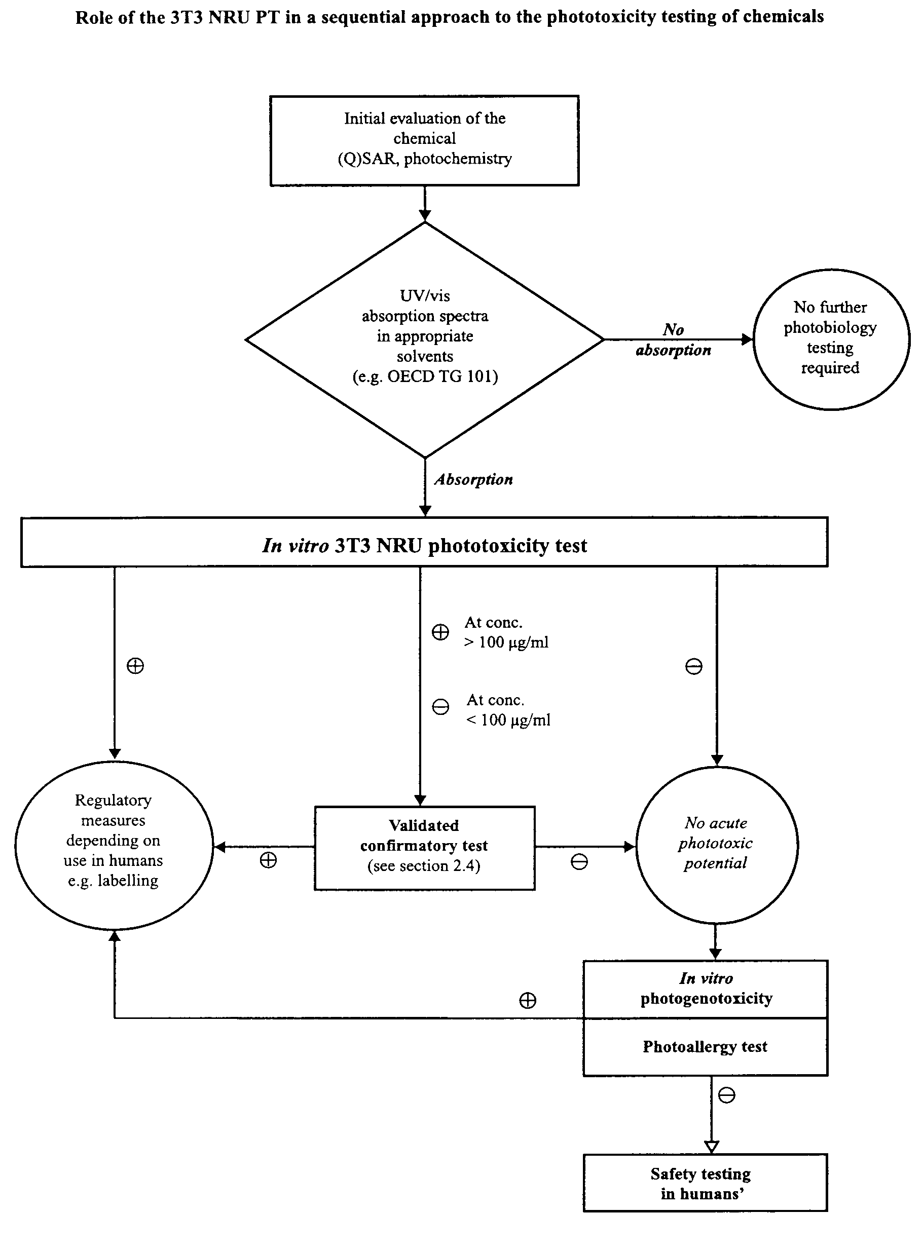 Official Journal of the European Communities