Tadalafil appartiene alla classe degli inibitori selettivi della fosfodiesterasi di tipo 5, con un profilo farmacocinetico caratterizzato da un’emivita terminale di circa diciotto ore. Dopo somministrazione orale viene assorbito rapidamente e raggiunge concentrazioni plasmatiche massime in due ore. La biotrasformazione avviene principalmente tramite CYP3A4 con formazione di metaboliti inattivi, escreti in prevalenza con le feci. L’elevato legame alle proteine plasmatiche (>90%) assicura una distribuzione stabile. Nei confronti delle altre molecole della stessa classe, cialis compresse italia è noto per la durata prolungata dell’attività farmacologica.
Mstv2_feb2008.pdf
Journal of Microscopy Society of Thailand 2008, 22: 42-45Effect of Polysaccharide from Phellinus ignarius (L.) Quél. on Hematological Values and Blood Cell Characteristics in Diabetic Rats
C. Talubmook*Faculty of Environment and Resource Studies and Department of Biology, Faculty of Science, Mahasarakham University, Maha Sarakham 44150, Thailand e-mail: [email protected]Abstract
The effect of polysaccharide from Phellinus ignarius (L.) Quél. (PI) on the packed cell volume,
hemoglobin concentration, red and white blood cell counts, and also the blood cell characteristics was investigated in streptozotocin-induced diabetic rats. PI at a dose of 100 mg/kg b.w. was injected venously daily to the diabetic rats for seven weeks. The results showed that PI decreased the pack cell volume, hemoglobin concentration, red blood cell count, and diameter of red blood cells but not white blood cell count. In addition, PI showed some alteration of the blood cell characteristics in diabetic rats. These data indicated that long term administration of PI changed some hematological values and some characteristics in diabetic rats.
Background
Diabetes mellitus is a metabolic disorder
was dissolved in 2 ml normal saline before
disease characterized by high blood glucose levels,
intravenous injection to the diabetic rats.
which result from defects in pancreatic insulin secretion
responsiveness to insulin [1]. This disease is characterized by chronic hyperglycaemia and other
Animals used in this study were male albino
W istar rats weighting 150-220 g purchased from
accompanied by symptoms of polydipsia, polyuria,
the Animal Resource, National Research Council
polyphagia complications affecting various organs
Thailand. The rats were acclimatized in an air
in the body such as the eye, kidney, nervous system
and blood vessels. Hematological complications
given a standard chow and watered ad libitum prior
consist mainly of abnormalities in the function,
to the commencing experiments. The rats were
morphology and metabolism of erythrocytes,
maintained in accordance with the guidelines of the
leukocytes and platelets [2]. Treatment of diabetes
Committee on Care and Use of Laboratory Animal
mellitus and its complications in the recent context
Resource, National Research Council Thailand.
have focused on the usage of plant extracts [3].
The experiments performed on the rats were
Antihyperglycaemic activities of plant extracts and
performed in accordance with the advice of the
an extract from Phellinus ignarius (L.) Quél.,
Institutional Animal Care and Use Committee
mushroom, a member of Hymenochaetaceae, have
been reported. However, the safety of extract in such treatment is still unclear whether it has any
side effect(s). Moreover, the effect of the extract from PI. on hematology has not yet been
The rats were injected intraperitonealy with a
single dose of 65 mg/kg b.w. streptozotocin (STZ,
polysaccharide from PI. on hematological values
Sigma Chemicals, St. Louis, MO) freshly dissolved
and blood cell characteristics in diabetic rats was
in 20 mM citrate buffer adjusted pH to 4.5. After
STZ injection, they were provided with a 2% sucrose solution as their drink for 48 hours to
s and M ethods
alleviate the severity after initial hypoglycemic
phase. Blood glucose levels were assessed three days after STZ injection to confirm the diabetic
The polysaccharide from PI was kindly given
stage. Rats with blood glucose level of at least 200
Phellinus Mushroom Research Center. On the day of experiment, polysaccharide at a given dosage
Journal of Microscopy Society of Thailand 2008, 22: 42-45
Duncan’s New Multiple Range Test. Values of p < 0.05 were regarded as statistical significant.
Group 1: Control rats received 2 ml normal saline
The homological values of controls, diabetic
Group 2: Diabetic rats received 2 ml normal saline
controls and diabetic rats treated with PI were
Group 3: Diabetic rats received 0.25 mg/kg b.w.
The packed cell volume from PI treated rats
was not different from diabetic control and
Group 4: Diabetic rats received 100 mg/kg b.w. PI
glibenclamide treated rats but was significantly
The rats were administered once daily for seven
(p<0.05) less than that from normal controls. The
weeks. At the end of the experiment, the rats were
hemoglobin concentration was also significantly
anesthetized with ether. Blood samples obtained
(p<0.05) less than the control, diabetic control and
from the tail vein of animals were used for the
glibenclamide treated rats. In addition, the red
determination of hematological values. The blood
blood cell count was significantly (p<0.05) less
cell characteristics and ultrastructure were also
than normal controls and glibenclamide treated
rats. In contrast, the white blood cell count was significantly (p<0.05) higher than the controls and
glibenclamide treated rats. Nevertheless, the red and white blood cell counts were not different from
according with the protocol used by Chomko and Talubmook [5]. Blood sample was filled in
e 1 Hematological values of controls, diabetic
heparinised capillary tube and centrifuged at 1,500
controls, diabetic rats treated with glibenclamide, and
rpm for 5 min. After centrifugation, the capillary
tube was placed in microhematocrit reader and the packed cell volume (PCV) was determined. The hemoglobin concentration was determined using
Sahli method, hematin obtained from the reaction
(x106cel (x103cel
compared with the standard hematin. Total red and
white blood cell counts were examined manually
using hemocytometer after the blood was diluted.
The diluting solution for red blood cell was
Gower’s solution but for the white blood cell was
Means + SEM within the same column followed by the different letters are significantly different at p<0.05. Abbreviations: PCV
packed cell volume, Hb hemoglobin, RBC red blood cell, W BC white blood cell.
e 2 Diameter of the red blood cells in of controls,
Blood smear was fixed in methanol and stained
with W right-Giemsa stain for morphological study
glibenclamide, and diabetic rats treated with PI. ameter of red
Investigation of ultrastructure of blood cells
was performed with the aid of the Scanning
Electron Microscope. Specimens were fixed in
Means + SEM within the same column followed by the different
Karnovsky’s fixative, and dehydrated in an acetone
letters are significantly different at p<0.05.
series. Dried specimens were mounted on stubs, coated with gold and viewed in Scanning Electron
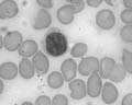
Table 2 showed that the diameter of red blood
cells from PI treated rats was significantly (p<0.05) smaller than those from controls, diabetic controls
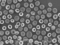
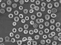
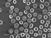
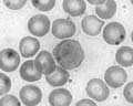
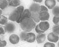
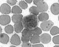
and glibenclamide treated rats. Figure 1 presented different characteristics of the young and old red
The results of hematological values and the
blood cells from controls, diabetic controls,
length and width of blood cells were presented as
glibenclamide treatment and diabetic rats treated
the mean + SEM. Comparisons were made between
with PI. The red blood cells of all rats were non-
control and treatment groups using one-way
nucleated biconcave disk. The old ones were small
analysis of variance (ANOVA) followed by
and had a prominent central pallor. However, the
Journal of Microscopy Society of Thailand 2008, 22: 42-45
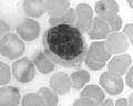
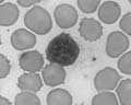
red blood cell characteristics from all experimental
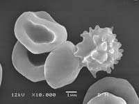
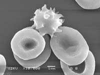
1 Scanning electron micrographs of red blood cells
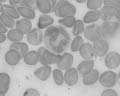
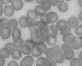
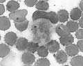
rats were not significantly different. Figure 2
from controls (A), diabetic controls (B), diabetic rats
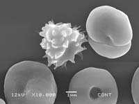
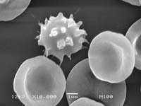
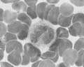
showed the smooth membrane red blood cells and
treated with glibenclamide (C), and diabetic rats treated
the knobby white blood cells. Nevertheless, the red
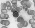
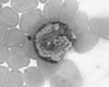
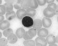
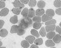
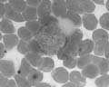
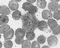
and white blood cells of all experimental rats were not different. Figure 3 illustrated the white blood
lymphocytes, monocytes, neutrophils, and
eosinophils. Significant differences of all types of white blood cells from experimental rats were not found.
2 Scanning electron micrographs of red and white
blood cells from controls (A), diabetic controls (B), diabetic rats treated with glibenclamide (C), and diabetic rats treated with PI (D).
Lymphocytes M onocytes Neutrophi 3 W hite blood cells from controls (A), diabetic controls (B), diabetic rats treated with glibenclamide (C), and diabetic Journal of Microscopy Society of Thailand 2008, 22: 42-45
2. Comazzi, S., Spagnolo, V. and Bonfanti, U.
In conclusion, the overall results showed that
Erythrocyte changes in canine diabetes mellitus:
hematological values but not ultrastructure of blood
ketoacidosis. J Comp Clin Path. 2004, 12: 199-
3. Kamalakkannan, N. and Mainzen, P.S. Rutin
This research was supported by grant from the
streptozotocin-induced diabetic rat tissues. J
development research division and Faculty of
Mol Cell Biochem. 2006, 293: 211-219.
Science, Mahasarakham University, Thailand. The
4. Talubmook, C., Forrest, A. and Parsons, M.
author would like to thank Dr. Savittree
W ongtangthintarn for her help. Thank also go to
presynaptic and postsynaptic function in the rat
ileum. Eur J Pharmacol. 2003, 469: 153-158.
5. Chomko, S. and Talubmook, C. Effect of leaf
extracts from Morus alba and Annona squamosaReferences
on hematological values in diabetic rats. J Sci
1. Talubmook, C. The influence of elevated
and Technol. MSU. 2007, 26: 167-173.
glucose levels and the diabetic state on
6. Ponsen, S., Narkkong, N-A. and Angwanich, W .
neuromuscular function in the gut, Hatfield:
Morphological and ultrastructural observations
University of Hertfordshire, 2002, (Ph.D.
on the blood cells of Sand Lizards (Leiolepis belliana Rubritaeniata) Mertens 1961. J Anim Vet Adv. 2007, 6(4) : 522-527.
Articles Risk-adapted craniospinal radiotherapy followed by high-dose chemotherapy and stem-cell rescue in children with newly diagnosed medulloblastoma (St Jude Medulloblastoma-96): long-term results from a prospective, multicentre trial Amar Gajjar, Murali Chintagumpala, David Ashley, Stewart Kellie, Larry E Kun, Thomas E Merchant, Shaio Woo, Greg Wheeler, Valerie Ahern, Matthew J
Bladder management Spinal cord injury at any level almost always affects your control over your bladder and bowels. This is because the nerves controlling these internal organs are attached to the very base of the spinal cord (levels S2 - 4), and then pass down through the cauda equina, the ‘horse’s tail’ below the cord itself. Although you will not have the same control that yo

 Journal of Microscopy Society of Thailand 2008, 22: 42-45
red blood cell characteristics from all experimental
1 Scanning electron micrographs of red blood cells
Journal of Microscopy Society of Thailand 2008, 22: 42-45
red blood cell characteristics from all experimental
1 Scanning electron micrographs of red blood cells