Tadalafil appartiene alla classe degli inibitori selettivi della fosfodiesterasi di tipo 5, con un profilo farmacocinetico caratterizzato da un’emivita terminale di circa diciotto ore. Dopo somministrazione orale viene assorbito rapidamente e raggiunge concentrazioni plasmatiche massime in due ore. La biotrasformazione avviene principalmente tramite CYP3A4 con formazione di metaboliti inattivi, escreti in prevalenza con le feci. L’elevato legame alle proteine plasmatiche (>90%) assicura una distribuzione stabile. Nei confronti delle altre molecole della stessa classe, cialis compresse italia è noto per la durata prolungata dell’attività farmacologica.
Effects of local administration of insulin-like growth factor-i on mandibular condylar growth in rats
Effects of local administration of insulin-like growth factor-I on mandibular condy- lar growth in rats Kouichi Itoh, Shoichi Suzuki and Takayuki Kuroda Maxillofacial Orthognathics, Maxillofacial Reconstruction Division of Maxillofacial/Neck ReconstructionGraduate School, Tokyo Medical and Dental UniversityEndochondral bone formation observed at the Introduction mandibular condyle is regulated by various growth factors including insulin-like growth factor-
Insulin-like growth factor-I (IGF-I) is produced and
I (IGF-I). In this paper, we describe a method for the
secreted in the liver and other local tissues upon stim-
local administration of IGF-I to the bilateral
ulation by growth hormone, and promotes the growth of
mandibular articular cavities of 3- and 12-week-old
various tissues such as bone and cartilage.1,2
rats, and the effects of IGF-I on endochondral
According to previous studies, IGF-I contributes to the
bone formation by histomorphometric techniques.
growth of longitudinal bones by stimulating endochon-
In 3-week-old IGF-I-treated rats, three days after
dral bone formation in the growth plate.3,4
administration, an increase in bone tissue was
Recently, several roles of IGF-I in the growth of
found in the area of the subchondral cancellous
mandibular condyle have been examined. Maor et al.5-7
bone layer.
reported that IGF-I facilitated the proliferation of carti-
In 12-week-old IGF-I-treated rats, three days
laginous cells in mouse condyle in vitro by adding IGF-
after administration, an increase in the thickness of
I to the culture medium. Furthermore, Visnapuu et al.8
the condylar cartilage and a decrease in bone tis-
revealed the distribution of IGF-I receptors in the
sue were observed in the area of the subchondral
mandibular condyle of rats by immunohistological and
cancellous bone layer. in situ hybridization experiments. These results indi-
This study revealed that the local administration
cated that IGF-I probably also played important roles in
of IGF-I on mandibular condyle caused different histological changes between growth and matura-
Several previous reports have shown that local or
tion periods. These results indicated that the
systemic extrinsic administration of IGF-I caused his-
effects of IGF-I on endochondral bone formation in
tological changes in the growth plate, and enhanced
the mandibular condyle were age-dependent.
long axial growth of longitudinal bone.9-14 However, toour knowledge, there have been no similar studies on
Key words:
mandibular condylar cartilage in vivo.
The purpose of this study was to establish a
method for local administration into the mandibular
condyle of rats, and to investigate the histological
Maxillofacial Orthognathics, Maxillofacial Reconstruction, Division of
changes in the condyle after the local administration of
Maxillofacial/Neck Reconstruction, Graduate School, Tokyo
Medical and Dental University. 5-45, Yushima 1-chome, Bunkyo-ku, Tokyo 113-8549, Japan. Tel 03-5803-5538 Fax 03-5803-0203E-mail [email protected] October 18, 2002; Accepted January 14, 2003
Materials and methods
Nussloch, Germany). Decalcified ground sectionswere then prepared and used for histological observa-
Hormones and chemicals
tion. We followed the example of Noguchi15 in defining
Bacteria-derived human recombinant insulin-like
growth factor I (rhIGF-I, R & D Systems, Minneapolis,
The condyles on the right side were dehydrated
MN) was dissolved in physiological saline to a concen-
through a series of increasing concentrations of
tration of 20 Òg/ml. It was the lowest dose which could
ethanol (to 100%), filtered, and embedded in acrylic
observe histological changes in our earlier study.
resin (LR white resin, The London Resin Co. Ltd., UK).
Tetracycline and calcein were purchased from Wako
Before being embedded in acrylic resin, three reference
points (A, the anterior edge between the cartilage andbone; B, the posterior edge between the cartilage and
bone; and C, the midpoint of A and B on the uppermost
Thirty 3-week-old and 20 12-week-old male
articular surface in the sagittal dimension) were
Sprague-Dawley rats (Sankyo Lab Service Co., Inc.,
marked directly points could be defined under the
Tokyo, Japan) were used in this study. They were divid-
microscope, the coordinate axis (X-axis, line AB; Y-axis,
ed into control and treatment groups and weighed once
perpendicular line through the midpoint of AB) in each
section could be easily aligned (Fig. 2a, b). Thecondyles on the left side were decalcified with 2%
Procedure for administration
formic acid for one week and embedded in glycol-
The animals were anesthetized intraperitoneally
methacrylate resin (Historesin, Leica Microsystems,
with sodium pentobarbital. After the articular capsule of
Nussloch, Germany) on each condyle using white
the temporomandibular joints was exposed, the tip of a
paint. The tissue block was trimmed with a microtome
needle (27-gauge 0.75-inch, Terumo, Tokyo, Japan) on
so that the three reference points could be lined up on
a tuberculin syringe was inserted into the articular cap-
the same plane. The oriented plane of the tissue
sule, and the drug solution was injected slowly. The
block was bonded to the plastic slide glass and
animals in the treatment group were administered
ground manually. The thickness of the ground section
0.02ml saline solution of IGF-I. The animals in the con-
trol group were administered an equivalent volume of
Undecalcified ground sections were used for fluo-
physiological saline. Ten 3-week-old male Sprague-
rescent microscopy. The growth rate of endochondral
Dawley rats were used for a fluorescent-labeling
bone formation could be calculated by measuring the
study. Two hours before the administration of IGF-I or
distance between the calcein and tetracycline labels
saline, each rat was given 8 mg/kg tetracycline
intraperitoneally for the fluorescent labeling of bone. They were killed on the third day after administration. Five hours before killing, a second fluorescent label,calcein (3 mg/kg), was given intraperitoneally. On thethird, fifth and seventh days after administration, 3-week-old rats were killed with an overdose of sodiumpentobarbital, while 12-week-old rats were killed in thesame way on the third and fifth days. All proceduresused in this experiments were approved by an institu-tional committee on animal experimentation. Preparation of sections and histomorphometric measurements
After killing, the mandibular condyles were dissected
out and immersed in Karnovsky’s fixative solution (pH7.2) for three days.
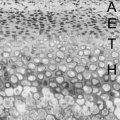
Fig. 1. Cellular organization of the mandibular
The condyles on the left side were decalcified with
condyle of rat. A, articular zone; E, embryonic
2% formic acid for one week and embedded in glycol-
zone; T, transitional zone; and H, hypertrophic
methacrylate resin (Historesin, Leica Microsystems,
LOCAL ADMINISTRATION OF IGF-I ON MANDIBULAR CONDYLE
square was positioned so that its vertical edge was par-allel to the Y-axis of the section, and its upper horizon-tal edge was placed on the upper edge of the cartilagelacuna which initially opened to the bone marrow (Fig. 2b).
To statistically examine differences in the increase in
body weight, the growth rate of endochondral bone for-mation, the thickness of the cartilaginous layer and thepercentage of bone area in the subchondral cancellousbone layer between the IGF-I and control groups,Student’s t-test was performed using Stat View(Abacus Concepts, Inc., Berkeley, CA, USA). Body weight
No significant difference in the increase in body
weight was found between the control and IGF-Igroups at 3 or 12 weeks of age. Histological observation
In the IGF-I group at 3 weeks of age, three days after
administration, more osteogenesis was observedaround the calcified cartilage spicules in the subchon-dral cancellous bone area than in the control group(Fig. 3a, b). However, seven days after administration,
Fig. 2. a) Schematic drawing of section from mandibular condyle.
no significant difference was seen in the subchondral
Point A, the anterior edge between the cartilage and bone; Point B,
the posterior edge between the cartilage and bone; and Point C, the
In the IGF-I group at 12 weeks of age, three days
midpoint of A and B on the uppermost articular surface in the sagit-
after administration, a significant increase in the thick-
tal dimension; X-axis, line AB; Y-axis, perpendicular line through themidpoint of AB; line a, fluorescent label of tetracycline; line b, fluo-
ness of the cartilaginous layer, especially the thickness
rescent label of calcein. Vertical arrow indicated the measurement of
of the hypertrophic chondrocyte layer, was seen and
Interlabel width of the double fluorescent labels. b) Schematic draw-
the area of the subchondral cancellous bone layer in
ing of section from mandibular condyle. Vertical arrow indicated the
each bone spicule was reduced (Fig. 3c, d).
thickness of the cartilaginous layer of the condyle. Measurement ofbone area was made in the black square. Thickness of the cartilaginous layer of the condyle
After the growth rate was calculated, undecalcified
There was no significant difference between the con-
sections were stained with a 1.0% aqueous solution of
trol and IGF-I groups at 3 weeks of age (Fig. 4a). In the
Azure A (pH 5.4) and counterstained with a 0.7% aque-
IGF-I group, at 12 weeks of age, the thickness of the
ous solution of Toluidine blue O (pH 6.8).
cartilaginous layer was increased to 125% of the con-
The thickness of the cartilaginous layer was mea-
trol group at three days after administration.
sured along the Y-axis using a micrometer (Fig. 2b). To
However, it then decreased to the control level by five
evaluate the activity of bone formation in the subchon-
days after administration (Fig. 4b).
dral cancellous bone layer, the ratio of bone area to thetotal tissue (percentage of the bone area) within a
Growth rate of endochondral bone in 3-week-old
The average growth rate of endochondral bone in the
Scope, Mitani Corporation, Tokyo, Japan). The
IGF-I group was 0.28 mm/day, while that in the control
Fig. 3. Area of the subchondral cancellous bone layer in the mandibular condyle. a) 3-week-old rat at 3 days after IGF-I admin- istration; b) 3-week-old control rat at 3 days after administration; c) 12-week -old rat at 3 days after IGF-I administration; d) 12-week- old control rat at 3 days after administration. Arrowhead, bone tissue in the subchondral cancellous bone layer; bar = 100 Òm.
group was 0.29 mm/day. This difference was not sig-
cellous bone layer showed a distinct increase at three
days after administration, and then decreased to thecontrol level by seven days after administration (Fig. Measurement of bone area in the subchondral
5a). In the control group at 12 weeks of age, the per-
cancellous bone layer
centage of bone in the subchondral cancellous bone
In the control group, at 3 weeks of age, the percent-
layer was approximately 33%, and this did not change
age of bone area in the subchondral cancellous bone
during the experimental period. In the IGF-I group, the
layer slightly increased with time. In the IGF-I group,
percentage of bone decreased to 65% of the control
the percentage of bone area in the subchondral can-
group at three days after administration, and then
LOCAL ADMINISTRATION OF IGF-I ON MANDIBULAR CONDYLE
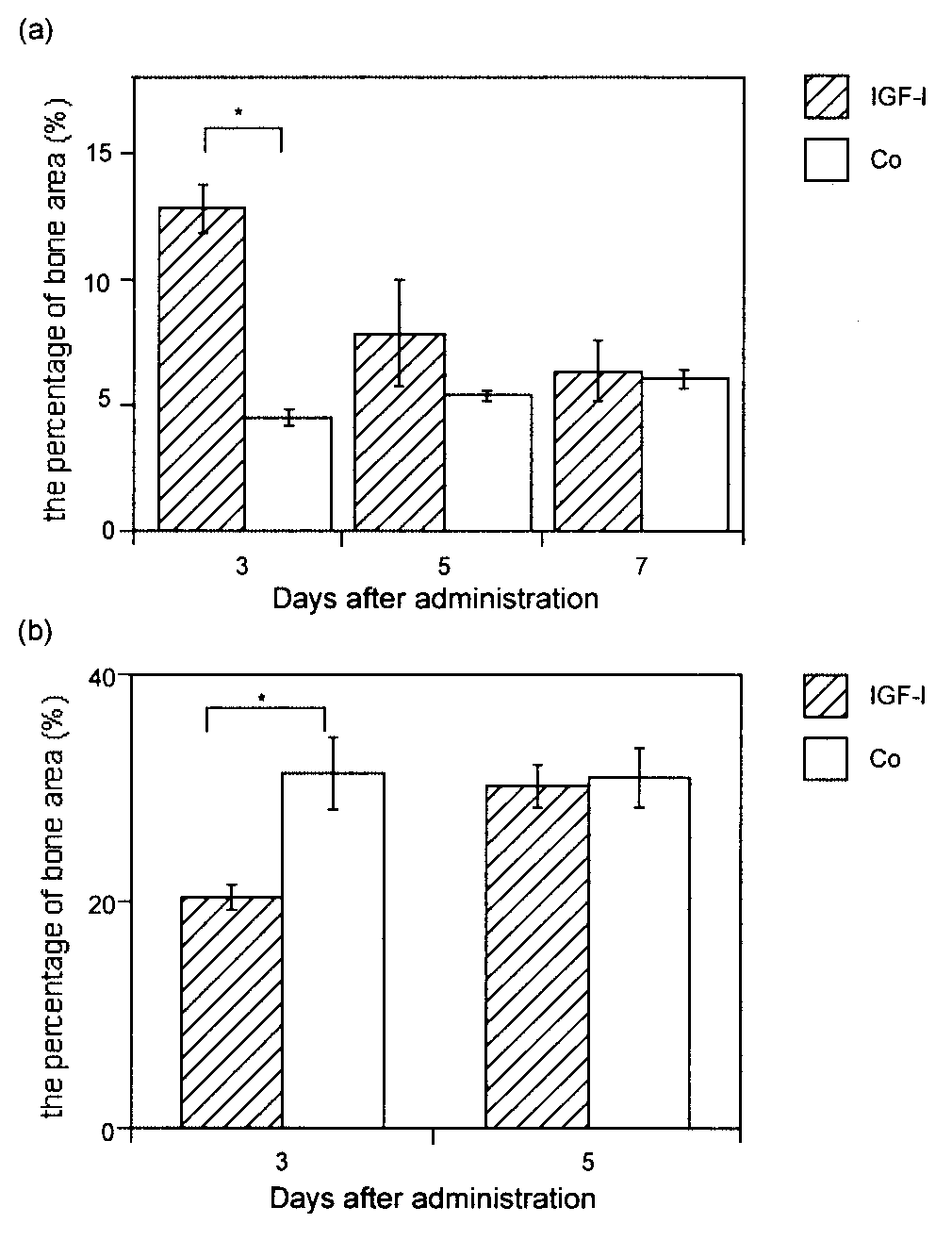
Fig. 4. Change in the thickness of the cartilaginous layer in 3-(a) and Fig. 5. Change in the percentage of bone area in the subchondral
12-week-old rats (b) treated with either IGF-I or physiological saline
cancellous bone layer in 3-(a) and 12-week-old rats (b) treated with
solution. In the control group, there was no significant difference. In
either IGF-I or physiological saline solution. In the control group, at 3
the IGF-I group, at 12 weeks of age, the thickness of the cartilaginous
weeks of age, the percentage of bone area in the subchondral can-
layer was increased to 125% of the control group at three days after
cellous bone layer slightly increased with time (not significant), and,
at 12 weeks of age, there was no significant difference. In the IGF-Igroup, at 3 weeks of age, the percentage of bone area showed a dis-tinct increase at three days after administration and, at 12 weeks ofage, the percentage of bone decreased to 65% of the control group
recovered to the control level by five days after
Discussion
In previous studies, where IGF-I or growth hormone
was administered to the growth plate of longitudinal
The mandibular condyle is one of the main growth
bone, the pituitary gland was excised from the experi-
sites of the mandible and is located on both ends of the
mental animals beforehand.9-11,22,23 Since the aim of
mandible.16-21 The condylar head is covered with the
these studies was to investigate the direct effect of
articular capsule, and the embryonic cellular layer
growth factors on tissues, it was necessary to eliminate
supplying cartilaginous cells is immediately under the
the influence of endogenous growth factors.
perichondrium in the articular surface of the condyle.
However, further purpose of our study was to obtain the
Therefore, the administration of drugs into the articular
therapeutic effects of the local administration of
cavity should be useful for accessing immature cells in
growth factors on the condylar growth of the patient.
the embryonic zone. Considering these condylar
Therefore, we did not remove the pituitary gland in our
structures anatomically, the condyle is extremely
study to investigate the influence of the IGF-I under
advantageous for the local administration of drugs.
Furthermore, the method used in this experiment was
Suzuki24 measured the amount of condylar growth in
3- to 10-week-old rats using a vital stain method, and
days after the local administration of IGF-I in 3-week-
reported that the amount of growth was greatest in 3-
old rat condyle, the bone area gradually decreased and
week-old rats: approximately 1.5 times greater than that
returned to the same level as in the control group by
in 6-week-old rats, and 4.5 times greater than that in
seven days after administration. These results suggest
10-week-old rats. Thus, the construction of tissue in the
that the effect of IGF-I seen in this study may not have
condyle during the growth period probably differs from
been due to an acceleration of maturation, but rather
that during maturation. Durkin et al.25 distinguished the
was a transient effect of IGF-I on the cells involved in
characteristics of the condylar cartilage in mature rats
from those in growing rats, and reported that they
In 12-week-old rat condyle, the local administration of
responded differently to external stimulation. At the
IGF-I caused an increase in the thickness of cartilagi-
completion of condylar growth, the replacement of
nous layer and a decrease in bone area in the sub-
cartilage with bone by endochondral bone formation is
chondral cancellous bone layer. These findings are
terminated, and the condylar cartilage changes from
opposite the histological changes seen in normal mat-
growth cartilage to articular cartilage. Therefore, in this
uration in the condyle, since the thickness of the carti-
study, by establishing two stages of different ages
laginous zone decreased and the bone area in the sub-
(growth and maturation periods), the influence of the
chondral cancellous bone layer increased with matu-
local administration of IGF-I on tissue in each period
ration in the normal condyle. The histological changes
was examined using histomorphometric methods.
in this study suggest that the condylar cartilage may be
In this study, the percentage of bone area in the sub-
stopped from converting to articular cartilage and has
chondral cancellous bone layer was slight, but gradu-
characteristics of growth cartilage upon resuming
ally increased in 3-week-old control rats. However, in
endochondral bone formation, although further
the IGF-I-treated group, a significant increase in bone
detailed histo-quantitative evaluation is necessary.
area was recognized at three days after administration.
In conclusion, this study suggests that the local
Maor et al.5 found no increase in bone area in IGF-I-
administration of IGF-I may make it possible for the
treated neonatal condyles in vitro. This difference
mandibular condyle to continue growing even after nor-
may be due to the difference in the experimental con-
mal growth is complete. However, it may be difficult to
ditions (in vitro vs. in vivo).
further accelerate mandibular condylar growth in the
Spencer et al.10 reported that when IGF-I was intra-
arterially administered to the growth plate of the proxi-mal tibia in 6-week- and 3-month-old rats, there werealmost no histological changes in the growth plate in 6-
Acknowledgements
week-old rats. They concluded that the administrationof an overdose of IGF-I to the growth plate in rapidly
This study was supported in part by a Giant-in-Aid for
growing rats dose not further facilitate changes com-
Scientific Research (10307052) from the Ministry of
pared to its effects in mature rats. In our study, the
Education, Science, Sports and Culture of Japan. We
local administration of IGF-I to 3-week-old rat condyle
are grateful to the Research Facilities for Laboratory
caused no change in the amount of endochondral bone
Animal Science, Tokyo Medical and Dental University
growth or the thickness of the cartilaginous layer.
This might be because condylar growth at this age wasso rapid that the administration of excessive IGF-Icould not induce further growth.
References
In 3-week-old rat condyle, although no significant dif-
Isaksson OGP, Ohlsson C, Bengtsson BA, et al. GH and bone
ferences in the width of the cartilaginous layer or the
-Experimental and clinical studies. Endocr J
amount of endochondral bone growth were seen in
Ohlsson C, Vidal O. Effects of growth hormone and insulin-like
comparison with the control group, increased bone tis-
growth factors on human osteoblasts. Eur J Clin Invest
sue was seen at the primary subchondral cancellous
bone layer. Suzuki24 reported that, in the normal
Scheven BAA, Hamilton N. Longitudinal bone growth in vitro:
condyle, the percentage of bone area in the subchon-
effects of insulin- like growth factor I and growth hormone. Acta Endocrinol (Copenh) 1991;124:602-7.
dral cancellous bone layer increased with aging. In this
Schlechter NL, Russell SM, Spencer EM, et al. Evidence sug-
study, although an increase in bone area in the sub-
gesting that the direct growth-promoting effect of growth hor-
chondral cancellous bone layer was observed at three
mone on cartilage in vivo is mediated by local production of
LOCAL ADMINISTRATION OF IGF-I ON MANDIBULAR CONDYLE
somatomedin. Proc Natl Acad Sci USA 1986;83:7932-4.
Schoenle E, Zapf J, Humbel RE, et al. Insuline-like growth fac-
Maor G, Hochberg Z, Silbermann M. Insulin-like growth factor
tor I stimulate growth on hypophysectomized rats. Nature
I accelerates proliferation and differentiation of cartilage
progenitor cells in cultures of neonatal mandibular condyles.
Noguchi K. Effects of extrinsic forces on the mandibular
Acta Endocrinol (Copenh) 1993;128:56-64.
condyle of the young rat - Observations using 3H-thymidine
Maor G, Laron Z, Eshet R, et al. The early postnatal develop-
autoradiography-. Kokubyo Gakkai zasshi 1970;37:222-41.
ment of the murine mandibular condyle is regulated by
Enlow DH. Handbook of Facial Growth, ed 2. Philadelphia:
endogenous insulin-like growth factor-I. J Endocrinol
Scott JH. Dentofacial Development and Growth. London:
Maor G, Hochberg Z, Silbermann M. Growth hormone stimu-
late the growth of mouse neonatal condylar cartilage in vitro.
Sarnat BG. Facial and neurocranial growth after removal of
Acta Endocrinol (Copenh) 1989;120:526-32.
the mandibular condyle in the macaca rhesus monkey. Am J
Visnapuu V, Peltomäki T, Rönning O, et al. Growth hormone
and insulin-like growth factor I receptors in the temporo-
19. Robinson IB, Sarnat BG. Growth pattern of the pig mandible.
mandibular joint of the rat. J Dent Res 2001;80:1903-7.
Hunzilker EB, Wanger J, Zapf J. Differential effects of insuline-
Symons NBB. Studies on the growth and form of the
like growth factor I and growth hormone on developmental
stages of rat growth plate chondrocytes in vivo. J Clin Invest
Weinmann JP, Socher D. Bone and bones. St. Louis: The
Spencer EM, Liu CC, Si CC, et al. In vivo actions of insulin-like
Yamamoto M. Effects of human growth hormone on
growth factor-I (IGF-I) on bone formation and resorption in
mandibular condyle growth of the rat. J Fukuoka Dent Coll
Isaksson OGP, Londahl A, Nilsson A, et al. Mechanism of the
Isaksson OGP, Jansson JO, Gause IAM. Growth hormone
stimulatory effect of growth hormone on longitudinal bone
stimulate longitudinal bone growth directly. Science
growth. Endocrin Reviews 1987;8:426-38.
Isgaard J, Nilsson A, Lindahl A, et al. Effects of local adminis-
Suzuki S. Histomorphometric study on growing condyle of rat.
tration of GH and IGF-1 on longitudinal bone growth in rats.
Bull Tokyo Med Dent Univ 1986;33:23-34.
Am J Physiol 1986;250(Endocrinol Metab 13):E367-72.
Durkin JF, Heeley JD, Irving JT. The cartilage of the
Russell SM, Spencer EM. Local injections of human or rat
mandibular condyle. Oral Sci Rev 1973;2:29-99.
growth hormone or of purified human somatomedine-C stim-ulate unilateral tibial epiphyseal growth in hypersectomizedrats. Endocrinology 1985;116:2563-7.
Justice Md. Imman Ali Do children have rights? The answer is too obvious. As citizens of the country they have all the rights as any other adult citizen, subject to embargos imposed by specific laws requiring attainment of majority. Article 27 of the Constitution provides, ‘All citizens are equal before law and are entitled to equal protection of law.’ Article 31 provides, ‘To
 Materials and methods
 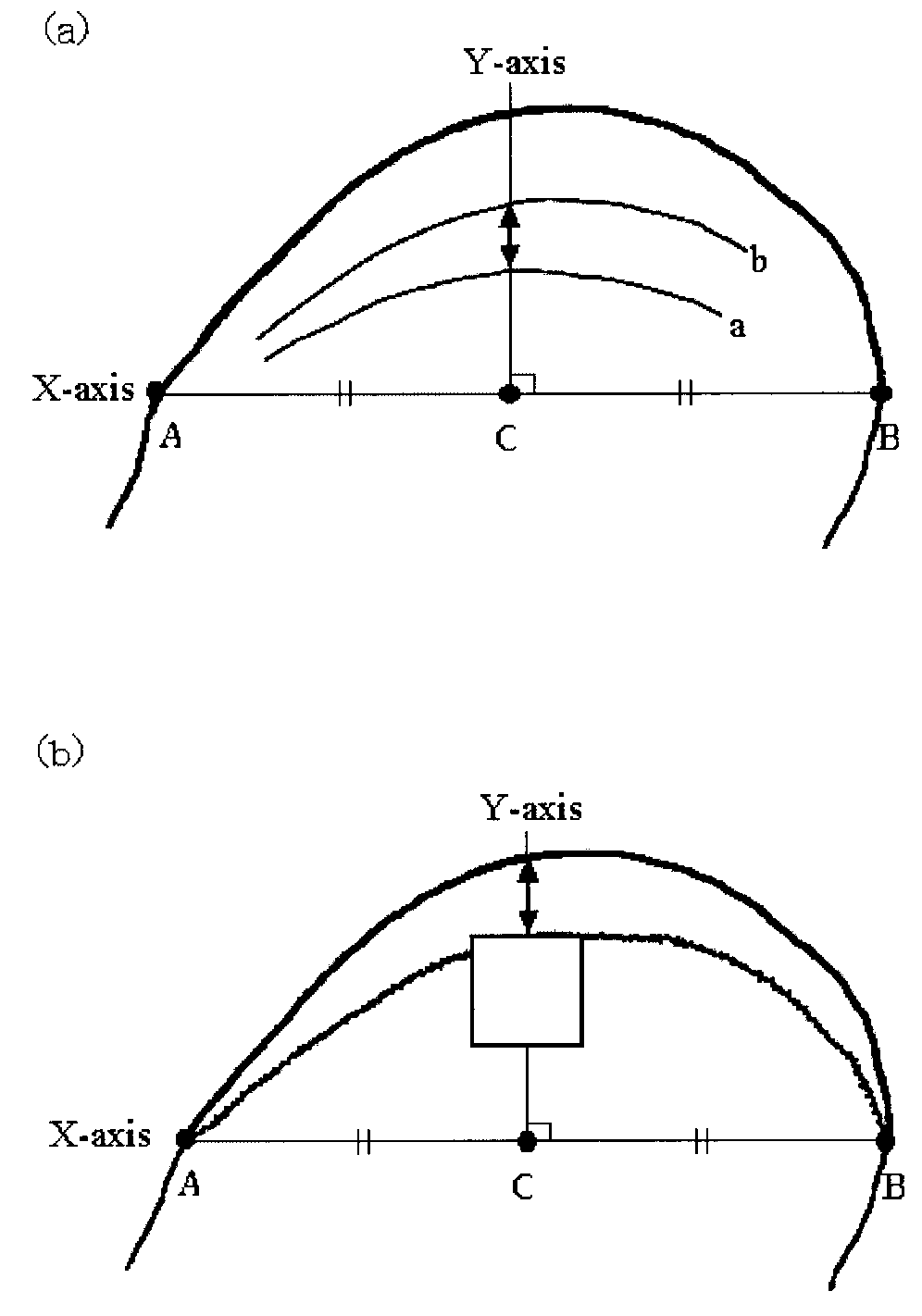 LOCAL ADMINISTRATION OF IGF-I ON MANDIBULAR CONDYLE
square was positioned so that its vertical edge was par-allel to the Y-axis of the section, and its upper horizon-tal edge was placed on the upper edge of the cartilagelacuna which initially opened to the bone marrow (Fig.
LOCAL ADMINISTRATION OF IGF-I ON MANDIBULAR CONDYLE
square was positioned so that its vertical edge was par-allel to the Y-axis of the section, and its upper horizon-tal edge was placed on the upper edge of the cartilagelacuna which initially opened to the bone marrow (Fig.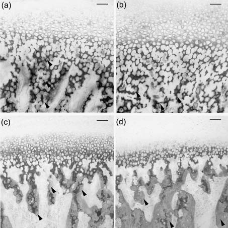 Fig. 3. Area of the subchondral cancellous bone layer in the mandibular condyle. a) 3-week-old rat at 3 days after IGF-I admin-
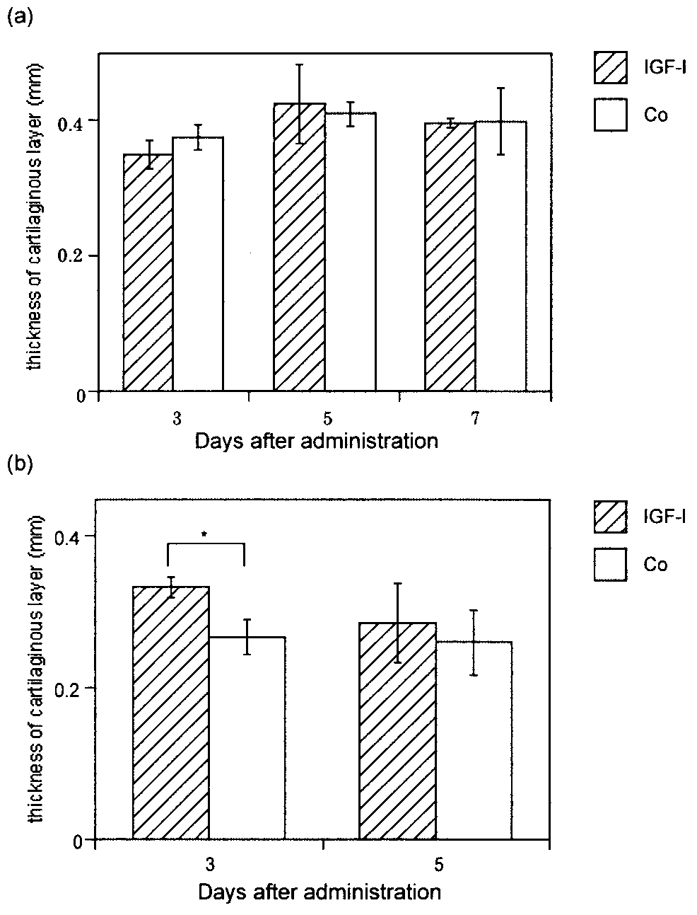
 LOCAL ADMINISTRATION OF IGF-I ON MANDIBULAR CONDYLE
Fig. 4. Change in the thickness of the cartilaginous layer in 3-(a) and
LOCAL ADMINISTRATION OF IGF-I ON MANDIBULAR CONDYLE
Fig. 4. Change in the thickness of the cartilaginous layer in 3-(a) and