Tadalafil appartiene alla classe degli inibitori selettivi della fosfodiesterasi di tipo 5, con un profilo farmacocinetico caratterizzato da un’emivita terminale di circa diciotto ore. Dopo somministrazione orale viene assorbito rapidamente e raggiunge concentrazioni plasmatiche massime in due ore. La biotrasformazione avviene principalmente tramite CYP3A4 con formazione di metaboliti inattivi, escreti in prevalenza con le feci. L’elevato legame alle proteine plasmatiche (>90%) assicura una distribuzione stabile. Nei confronti delle altre molecole della stessa classe, cialis compresse italia è noto per la durata prolungata dell’attività farmacologica.
Microsoft powerpoint - ppt0000018 [read-only]
Investigation of Ibuprofen release from PVP, PVP/alginate and PEO/alginate nanofibers.
A.Y.A Kaassis, Nicholas P. Chatterton, Gareth R.Williams Faculty of Life Sciences, London Metropolitan University, 166-200 Holloway Road,INTRODUCTION
Electrospinning is an effective technique in the generation of polymeric fibers with a diameter in
the range of a micro to nanometre, through a contactless process employing electric fields (Li &
Xia 2004). Electrospun nanofibers have attracted much research attention for several applications.
Energy storage is one purpose, where nanofibers have been used as a storage device for
alternative energy sources such as hydrogen and natural gases. Secondly, fibers may be used for
ecological and biomedical purposes, such as sensors for chemical and biochemical detection.
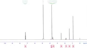
Figure 2: FTIR spectra of (pink) ibuprofen; Figure 3: The 1H NMR spectrum of PEO-SA with
Moreover, nanofibers are able to contribute to various medical areas including drug delivery,
(Black) PEO-SA-IBU and (pink) PEO-alg. sodium ibuprofen in D O.
genomic medicine, scaffolds for tissue engineering and smart wound dressings (Young-Seak & Ji
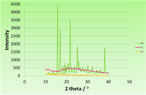
XRD patterns, given in Figure 4, showed that none of crystalline peaks of the drug were
exhibited by the fibers, suggesting that ibu exists in an amorphous form in the fibers, and that the
Ibuprofen is a non-steroidal anti-inflammatory drug, frequently used in the treatment of
fiber components are mixed at the molecular level.
osteoarthritis, rheumatoid arthritis and inflammation. It also has analgesic properties. However,
its long-term administration causes gastrointestinal side effects such as ulceration, bleeding and
perforation. Ibuprofen has a short biological half-life of 2.0 ± 0.5 hours. Therefore, repeated doses
are necessary to maintain therapeutic efficacy over long periods of time (Kachrimanis et al.
The purpose of this study was to encapsulate ibuprofen (ibu) into electrospun nanofibers using
different polymer carrier materials, and to investigate their potential as prolonged release systems
in different milieus. Polyvinylpyrrolidone-ibuprofen (PVP-ibu), Polyvinylpyrrolidone/alginate-
ibuprofen (PVP-SA-ibu), and poly (ethylene oxide)/alginate ibuprofen (PEO-SA-ibu) fibers were
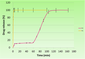
Figure 4: XRD patterns: sodium ibuprofen (IBU); Figure 5: Drug release from PEO-SA –IBU nanofibers
generated by electrospinning and characterised by FT-IR, scanning electron microscopy (SEM)
(N2) and PVP-IBU nanofibers (N1) in acetate buffer
Differential scanning calorimetry was additionally used to characterise the fibers, and supported
the molecular dispersion of ibu in the fibers. 1H NMR spectra of fibers dissolved in D O
Ibuprofen loaded fibers were prepared by using 10% (w/v) PVP 360 (Mw = 36 000 Da), 10 %
(figure 3) showed that structure of ibu was preserved after electrospinning, and not altered by the
PVP / 2% SA (Sodium alginate) and 10% PEO (Polyethylene oxide) (Mw = 40.000 Da) / 2% SA
as carrier polymers. 1% (w/v) drug polymer solutions used for spinning. Electrospinnning was
A dissolution study in an acetate buffer, the results of which are given in Figure 5, showed that
performed with a flow rate of 0.3 ml/h, a spinnaret to distance of 15 cm, and an accelerating
the release of ibu from PVP nanofibers was very fast. The PVP-SA fibers released more slowly,
and the PEO-SA nanofibers displayed the slowest release. The PVP nanofibers dissolved within
The morphology and structures of nanofibers were studied by scanning electron microscopy
30s and released all the encapsulated drug. The PVP-SA nanofibers liberated 100% of the
(SEM) and X-ray diffraction (XRD). The stability of Ibuprofen during the electrospinning process
incorporated drug after 5min. The PEO-SA nanofibers floated for one hour then sank and started
was assessed using Fourier transform infrared spectroscopy (FTIR) and nuclear magnetic
dissolving. During the first 60 min the drug release rate was slow and reached a plateau at
resonance (NMR). In vitro release of ibuprofen from the fibers was carried out at 37 ºC in three
13.6 %. After 60 min, drug release accelerated and peaked after 127 min, where 100% of the drug
media: an acetic acid buffer (pH 3), simulated intestinal fluid (pH 6.8) and phosphate buffered
had been released. The drug release was faster in SIF. The PVP nanofibers dissolved entirely with
saline (pH 7.4). Drug concentrations in solution were measured by UV spectroscopy. All
one minute. After 10 min, 60 % of the drug was released from the PEO-SA nanofibers;
measurements were carried out in triplicate.
nevertheless, more than 40% of the drug load stayed in the nanofibers. CONCLUSION
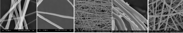
SEM images (Figure 1) showed that all the fibers with had smooth surfaces and cylindrical
In conclusion, sodium ibuprofen was successfully loaded into nanofibers without altering its
shapes. The fiber diameters for pure PVP and PVP-ibu fibers were around 450nm. SEM images
structure, as proven by UV spectra, FTIR and NMR. The fibers were found to be uniform in size
(Figure 1) showed that all the fibers with had smooth surfaces and cylindrical shapes. The fiber
and shape. Drug release from the fibers was studied in realistic in vitro conditions, and an
diameters for pure PVP and PVP-ibu fibers were around 450nm.
interesting two-stage release mechanism observed for PEO-SA-ibu fibers.
REFERENCES
Kachrimanis, K., Nikolakakis, I. & Malamataris, S., 2000. Journal of Pharmaceutical Sciences, 89(2), pp.250-9. (a) (b) (c)
Li, D. & Xia, Y., 2004. Advanced Materials, 16(14), pp.1151-1170.
Figure 1: SEM images of the nanofibers. a) PVP; b) PVP-Ibu; c) PEO-SA-Ibu; d) PVP-SA; e) PVP-SA-Ibu.
Young-Seak, L. & Ji Sun, I., 2010. Preparation of Functionalized Nanofibers and Their Applications. In K. Ashok, ed. Nanofibers. India: InTech, pp. 121-138.
FTIR spectra (figure 2) confirmed successful incorporation of ibu into the fibers, but also showed
ACKNOWLEDGEMENTS
small shifts in the positions of the characteristic absorbances. The peaks shift suggest that
We thank the EPSRC and Queen Mary, University of London, for access to SEM facilities, and
interactions such as H-bonding existed between ibuprofen and the polymers.
AYK thanks London Metropolitan University for provision of a PhD studentship. RESEARCH POSTER PRESENTATION DESIGN 2011 www.PosterPresentation
Land Use, Residential Density, and Walking The Multi-Ethnic Study of Atherosclerosis Daniel A. Rodríguez, PhD, Kelly R. Evenson, PhD, Ana V. Diez Roux, MD, PhD, Shannon J. Brines, MS Background: The neighborhood environment may play a role in encouraging sedentary patterns,especially for middle-aged and older adults. The aim of this study was to examine the associations between walking an
Gesammelte Vitamin E Studien Vitamin E schützt die Blutgefäße Raucher nehmen beim Inhalieren einer Zigarettebüberdurchschnittlich viele freie Radikale auf,welche die Peroxidation atherogener Lipoproteine begünstigt.Vitamin E als potenter Radikalfänger wirkt als diesen Vorgängen entgegen. Fazit der Studie: Die Supplementierung von 200mg/d Vitamin E schützt deutlich vor der Peroxid


 Investigation of Ibuprofen release from PVP,