Tadalafil appartiene alla classe degli inibitori selettivi della fosfodiesterasi di tipo 5, con un profilo farmacocinetico caratterizzato da un’emivita terminale di circa diciotto ore. Dopo somministrazione orale viene assorbito rapidamente e raggiunge concentrazioni plasmatiche massime in due ore. La biotrasformazione avviene principalmente tramite CYP3A4 con formazione di metaboliti inattivi, escreti in prevalenza con le feci. L’elevato legame alle proteine plasmatiche (>90%) assicura una distribuzione stabile. Nei confronti delle altre molecole della stessa classe, cialis compresse italia è noto per la durata prolungata dell’attività farmacologica.
Powerpoint presentation
What are they really doing in there?
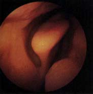
The nose contains three major structures which are key to ordinary breathing. They are:
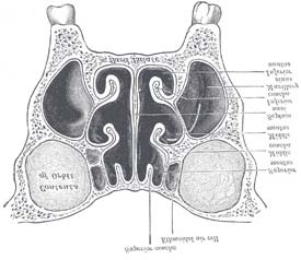
Sinuses are air-filled cavities in the skull, around the nose.
On each side of the face, there are four sets of sinuses: Frontal – above the eyes Maxillary – in the cheek bones Ethmoid* – behind the nasal bridge Sphenoid – deep behind the eyes
All sinuses are connected to the inside of the nose through an opening called an ostium.
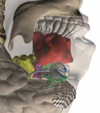
The basic concepts of endoscopic sinus surgery are fairly straightforward. However, to follow the complex steps of the procedure, in-depth understanding of nasal sinus anatomy is essential.
9 Use multiple codes for multiple sinuses
± Don’t code a diagnostic endoscopy with surgical
± Don’t code removal of the uncinate process separately
± Don’t routinely code turbinectomy separately
~ Do code turbinectomy separately when there’s pathology
~ Do code septoplasty separately
Some nasal sinus endoscopies use computer-assisted navigation.
A special headset is placed on the patient’s head. This relays data to a computer builds a 3-D model the surgeon uses to map out the anatomy in advance. The computer also produces 3-D images of the procedure in progress, essentially allowing the surgeon to see around corners.
Computer-assisted navigation is most common in patients whose anatomy has been altered, either by severe disease or by previous sinus procedures.
Non-Endoscopic Sinus Surgery:Frontal Sinus Trephination
In addition to FESS, the frontal sinus can also be accessed by simple frontal trephination.
An incision is made under the brow and a hole is drilled into the frontal sinus. An external drain is then placed through the hole for irrigation and sinus drainage.
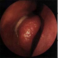
The patient was prepped and draped for endoscopic sinus surgery. The patient’s right nasal cavity was injected with 1% lidocaine with epinephrine. Under 0 degree endoscopic visualization, a sickle knife was used to make an uncinectomy and the middle turbinate was medialized. Straight and upbiting forceps were used to takedown the bulla and anterior ethmoid cells. A 30 degree endoscope was then used to open up the maxillary sinus widely using backbiting forceps and a straight Allen. The nasal cavity was suctioned clear of blood. Bactroban-coated sponge was placed in the dissected ethmoid air cells and secured to the side of the face with steristrips. The patient left the OR in satisfactory condition.
For the complete version of this topic or for more information on any other topics, contact us at :
or e-mail us at : [email protected]
Case 2:10-cv-01736-DSC-RCM Document 88 Filed 01/19/12 Page 1 of 11 IN THE UNITED STATES DISTRICT COURT FOR THE WESTERN DISTRICT OF PENNSYLVANIA REPLY BRIEF IN SUPPORT OF DEFENDANTS’ MOTION FOR CASE MANAGEMENT ORDER [ORAL ARGUMENT SCHEDULED] Case 2:10-cv-01736-DSC-RCM Document 88 Filed 01/19/12 Page 2 of 11 INTRODUCTION Plaintiffs oppose Defendants’ Motion for Case Manag
ASAMBLEA GENERAL EXTRAORDINARIA “CLUB RADIOCONTROL VIÑA DEL MAR” ACTA N° 03/2011 En Viña del Mar, a 11 de Febrero del año 2011, siendo las 21.20 horas, en la calle Coraceros N° 50 de esta Comuna, Provincia de Valparaíso, Región Vta., se lleva a efecto la Asamblea General Extraordinaria de Socios, con las personas que se individualizan y firman el Anexo de Asistencia de S
 What are they really doing in there?
 The nose contains three major structures which are key to ordinary breathing. They are:
 Sinuses are air-filled cavities in the skull, around the nose.
On each side of the face, there are four sets of sinuses: Frontal – above the eyes Maxillary – in the cheek bones Ethmoid* – behind the nasal bridge Sphenoid – deep behind the eyes
All sinuses are connected to the inside of the nose through an opening called an ostium.
Sinuses are air-filled cavities in the skull, around the nose.
On each side of the face, there are four sets of sinuses: Frontal – above the eyes Maxillary – in the cheek bones Ethmoid* – behind the nasal bridge Sphenoid – deep behind the eyes
All sinuses are connected to the inside of the nose through an opening called an ostium.
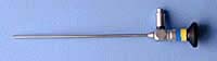
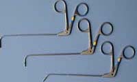
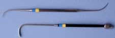
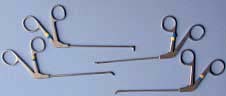
 The basic concepts of endoscopic sinus surgery are fairly straightforward. However, to follow the complex steps of the procedure, in-depth understanding of nasal sinus anatomy is essential.
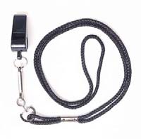
 9 Use multiple codes for multiple sinuses
± Don’t code a diagnostic endoscopy with surgical
9 Use multiple codes for multiple sinuses
± Don’t code a diagnostic endoscopy with surgical 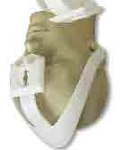
 Some nasal sinus endoscopies use computer-assisted navigation.
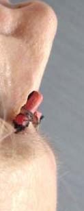
 Non-Endoscopic Sinus Surgery:Frontal Sinus Trephination
In addition to FESS, the frontal sinus can also be accessed by simple frontal trephination.
An incision is made under the brow and a hole is drilled into the frontal sinus. An external drain is then placed through the hole for irrigation and sinus drainage.
Non-Endoscopic Sinus Surgery:Frontal Sinus Trephination
In addition to FESS, the frontal sinus can also be accessed by simple frontal trephination.
An incision is made under the brow and a hole is drilled into the frontal sinus. An external drain is then placed through the hole for irrigation and sinus drainage. The patient was prepped and draped for endoscopic sinus surgery.
 For the complete version of this topic or for more information on any other topics, contact us at :
or e-mail us at :
For the complete version of this topic or for more information on any other topics, contact us at :
or e-mail us at :